
Introduction: Type 1 diabetes mellitus (T1DM) is a disease characterized by the destruction of the insulin-producing, pancreatic β-cells, where the inability to maintain glucose homeostasis causes multi-organ problems. The influence of T1DM on exercise performance is complex, with some studies displaying no difference (Baldi et al., 2010) and others finding an impaired exercise capacity in T1DM compared to healthy individuals (Goulding et al., 2020). When rigorous matching of adults with T1DM for age-, sex- and physical activity to healthy controls is performed, however, the balance of evidence indicates a reduced exercise capacity in T1DM (Eckstein et al., 2020). The cause of this lower exercise capacity seems to be related to an impaired mitochondrial function in individuals with T1DM irrespective of physical activity (Monaco et al., 2018). Whether skeletal muscle mitochondria of people with T1DM are less responsive to exercise training has not been systemically studied.
Objectives: To compare aerobic exercise capacity and mitochondrial function between adults with T1DM and matched adults without T1DM both before and after an aerobic exercise training intervention.
Methods: Data collection is still ongoing, but so far 17 adults with T1DM and 5 matched healthy control subjects (18-65 years) have performed a maximal ramp incremental test on a cycle ergometer, before and after a 4-week moderate-intensity aerobic exercise training intervention. Muscle biopsies were obtained from the vastus lateralis before and after the intervention. Mitochondrial respiration was assessed in permeabilized fibres using high-resolution respirometry and transmission electron microscopy was to study mitochondrial ultrastructure.
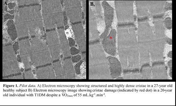
Results: The current low sample size and unequal groups preclude statistical analysis at present. Maximal oxygen uptake (VO2peak) was 37±15 vs. 47±11 mL.kg-1.min-1 for people with T1DM (n=17, 40±17 yrs) and controls (n=5, 30±15 yrs). Maximal OXPHOS capacity in permeabilized fibers was 80±44 vs. 98±28 pmol.s-1.mg-1 in T1DM and individually-matched controls, respectively (i.e., n=5 for both groups). Peak power (pre: 279±81 vs. post: 292±96 W), VO2peak (pre: 40±13 vs. post: 41±13 mL.kg-1.min-1) and OXPHOS capacity (pre: 83±26 vs. post: 107±44 pmol.s-1.mg-1) have been assessed before vs. after training in a subgroup of 5 people with T1DM, with matched controls currently undergoing the same intervention. Moreover, we observed mitochondrial cristae damage in electron microscopy images from one healthy, and moderately-trained (VO2peak 55 mL.kg-1.min-1), person with T1DM (Figure 1).
Conclusion: These data will allow us to comprehensively study mitochondrial alterations and mechanisms underpinning the impairment in exercise tolerance in people with T1DM. Moreover, this study will reveal to whether skeletal muscle mitochondria of people with T1DM show plasticity after aerobic exercise training. It is expected that data collection and analysis is complete mid-April 2022.
Biomedical Basis of Elite Performance 2022 (University of Nottingham, UK) (2022) Proc Physiol Soc 49, PC21
Poster Communications: Influence of aerobic training on exercise capacity and mitochondrial function in type 1 diabetes and matched healthy controls
Richie Goulding1, Matthijs van der Laan1, Anne Strating1, Wendy Noort1, Aryna Kolodyazhna1, Frank Bloemers2, Rob Wüst1
1 Vrije Universiteit 2 Amsterdam UMC
View other abstracts by:
Where applicable, experiments conform with Society ethical requirements.