Introduction
Affecting over 20 million people worldwide, kidney diseases are primarily caused by nephron lesions. The complex tubular structure of nephrons challenges accurate diagnosis through a kidney biopsy. To address this, we developed a workflow combining optical clearing and artificial intelligence, enhancing accuracy and automating the process. Optical clearing turns samples transparent, allowing precise mapping of internal tubular configurations. Light-sheet microscopy images these structures, but accurate segmentation is necessary for quantifying measurements, such as tubular length and volume. With its remarkable capabilities in medical imaging analysis, machine learning presents a promising solution for the tubule segmentation task. We propose a vision transformer-based machine learning segmentation model that automates measurements, aids clinicians in generating appropriate diagnoses, and provides valuable insights into gene location and function. This highly translational method can be extended to other tubular organs, benefiting a broader community by quickly adapting the underlying code.
Methods
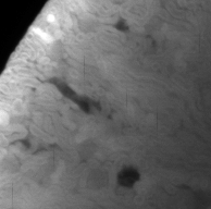
We collected 36 light-sheet microscopy images, each representing a quarter of a mouse kidney. These 16-bit, 3-channel images have 2048×2048 pixels, with a depth range (Z) of 700-1200 pixels and pixel sizes of 4.08um (XY plane) and 4.0um (Z axis). Due to limited RAM and VRAM, images are split into fixed-size blocks and segmented individually. Each block is treated as a series of 2D images. Except for the 1st image, a convolutional neural network encodes and captures local features of the Nth image and the segmentation result of the (N-1)th image together, as adjacent images usually share spatial information. The transformer encoder learns high-level contextual representations while the transformer decoder reconstructs the structure. The convolutional decoder recovers spatial dimensions and generates a tubule probability map, with the final segmentation mask obtained by normalizing the output probability map using a softmax activation function. At last, these segmented blocks are reconstructed into the original size image as the segmentation result.
Results and Discussion
While the samples have been annotated, we are still in the process of building the model. Preliminary tests (Classical 3D Convolutional Neural Network) demonstrate the potential of the proposed approach to effectively segment tubular structures in the kidney, with initial results indicating improvements in segmentation accuracy compared to traditional methods. Upon completion of the model, we will conduct a comprehensive evaluation, including quantitative metrics such as Dice score, precision, recall, and intersection over union (IoU), to assess the model's performance on the segmentation task. Additionally, we will investigate the impact of incorporating different pre-processing techniques, data augmentation strategies, and loss functions to enhance the model's accuracy and generalization further. Comparisons with existing segmentation methods, both machine learning-based and conventional, will establish the superiority of our proposed approach. Finally, we plan to analyze the model's performance in real-world clinical settings, exploring its potential for assisting clinicians in making accurate diagnoses and offering valuable insights for researchers in studying gene location and function in 3D spatial contexts.