Diabetic Cardiomyopathy (DCM) is a subset of heart failure that is a consequence of diabetes mellitus1. The contractile dysfunction of heart failure is characterised by blunted beta-adrenergic-cAMP signalling2. Insulin is known to reduce cAMP via phosphodiesterase (PDE)3, which experiences dysfunction in the diabetic state, owing to insulin resistance. Recently insulin and Fibroblast Growth Factor 1 (FGF1) signalling has been shown to be convergent4 and appears to be protective against DCM5. We tested whether cAMP signalling is impaired in a model of DCM using human induced pluripotent stem cell (hiPSC) derived cardiomyocytes, and investigated whether cAMP kinetics is altered by FGF1.
Healthy hiPSC (OX1-19, obtained from The James Martin Stem Cell Facility, Oxford) were differentiated into cardiomyocytes and cultured in either normal or high-glucose, high-fatty acid media. Cytoskeletal and nuclear staining were used to assess cell and nuclear area. ATP based cell viability and glucose reuptake were assessed using a luminescence-based assay. BNP release was measured by a NT-proBNP ELISA kit and gene expression levels were detected by bulk RNA-sequencing. Real time cAMP kinetics were investigated with a fluorescence resonance energy transfer (FRET) based sensor Epac-SH187. DCM model cells expressed phenotypic changes consistent with the disease. Cells cultured in either 11 mM (n=122 cells, 2 wells) or 25 mM glucose (n=102 cells, 2 wells) had a significant increase in cell area (µm2) compared to control (n=236 cells, 2 wells), (p<0.0001, Kruskal-Wallis). DCM cells (n=7 separate wells) had a significant increase in the concentration of NT-proBNP, a key hypertrophic marker, in the media assay compared to control cells (n=7 separate wells, p<0.0001, one-way ANOVA). DCM cells (n=7 separate wells) had a significantly lower cell viability compared to control (n=7 separate wells, one-way ANOVA, p<0.001). RNA-seq informed the KEGG pathway analysis were enriched in PI3-Akt, cAMP signalling, Ras-Raf (ERK-MAPK), and hypertrophic cardiomyopathy pathways. Hypertrophic genes, collagens, and ANP and BNP genes, both of which are released in failing heart, were upregulated, as was PDE4DIP. cAMP signalling in response to forskolin was lowered in the DCM model cells (n=21, 7.21±0.05%) compared to control (n=20, 9.53±0.12%, p<0.05). This was reversed when DCM cells were treated with FGF1 (1 ng/ml, n=19, 13.65±0.09%, p<0.0001).
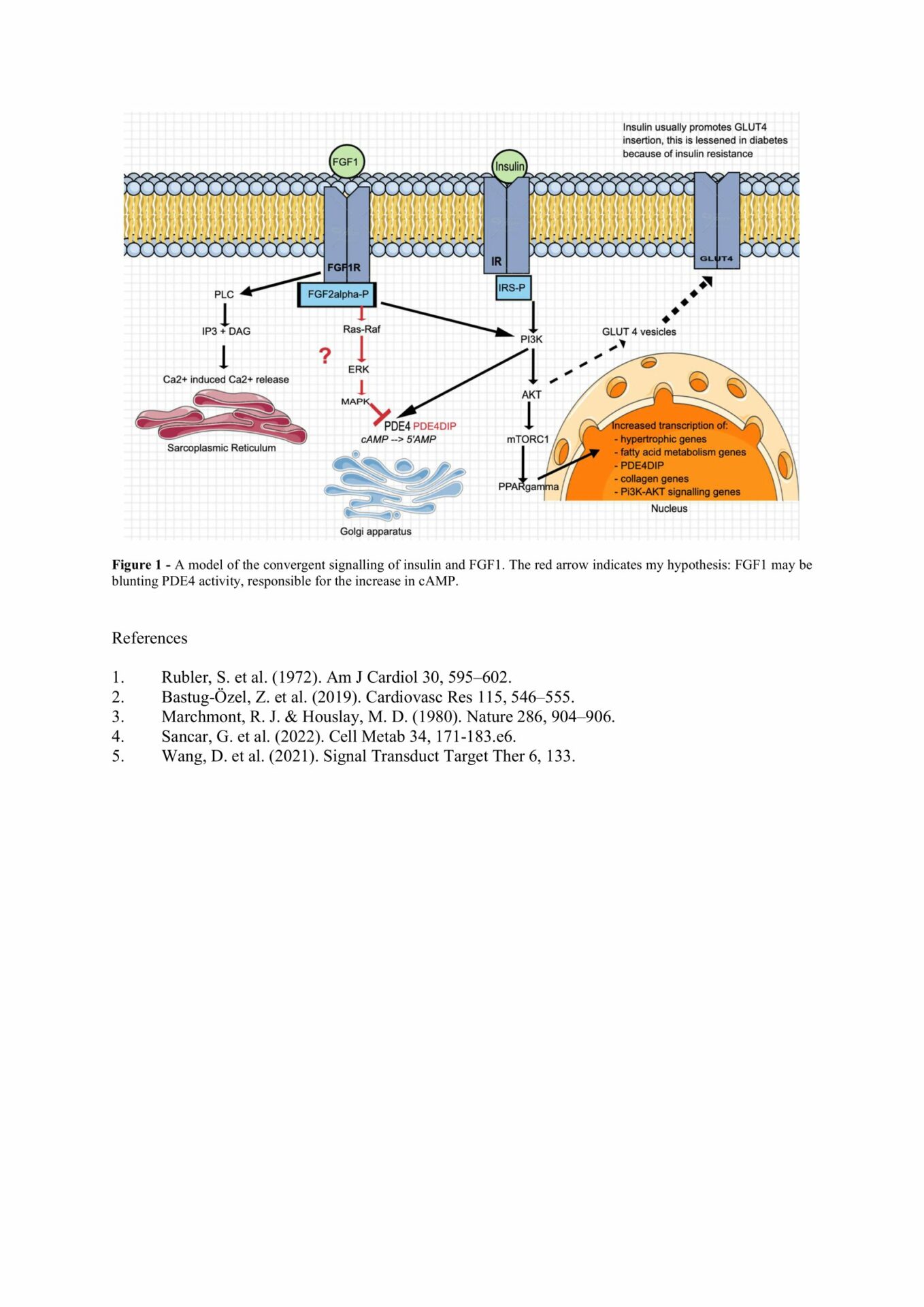
hiPSC-derived cardiomyocytes cultured in high-glucose, high-fatty acid media produce characteristics of DCM. cAMP signalling was blunted in DCM cells. This response was ameliorated by increasing FGF1. Gene expression changes point to a novel type of FGF1 signalling pathway in the heart, which might lead to improved contractility, as postulated in Figure 1.
Figure 1 – A model of the convergent signalling of insulin and FGF1. The red arrow indicates a hypothetical pathway where FGF1 may be blunting PDE4 activity that is responsible for the increase in cAMP.