Introduction: The hamstring muscles are a complex group with distinct architectural characteristics (Kellis, 2018). Surface electromyographic (EMG) signal amplitude is typically used to compare the neural drive to hamstrings muscles but previous studies have documented conflicting results regarding their activation during knee flexion across different knee joint angles (Kellis & Blazevich, 2022).
Aims: The purpose of the present study was to examine the differences between the Biceps femoris (BF) and Semitendinosus (ST) contribution at two different knee joint angles (0° = full extension, and 90°).
Method: Fifteen healthy active participants performed isometric knee flexions at four target forces [10%, 20%, 40%, and 60% of Maximum Voluntary Contraction (MVC)] at two knee joint angles. High-density EMG was used to examine the motor unit (MU) discharge characteristics (mean discharge rate, coefficient of variation of interspike interval and standard deviation of filtered cumulative spike train) from the BF and ST. Linear mixed effects models were applied to compare the MU discharge characteristics between the two muscles (BF and ST), knee angles (0° and 90°) and target forces (10%, 20%, 40%, and 60%). In all models, muscles, knee angle and force level were specified as fixed factors with participant as a random factor.
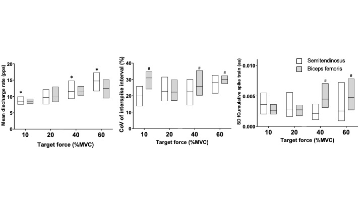
Results: The MVC force was greater at the 0° knee angle (long length) compared to 90° (p = 0.002). Additionally, the coefficient variation of force was greater at 90° knee angle compared to 0° (p = 0.032). The mean discharge rate was greater for the ST compared to the BF (p = 0.022). However, the variability in discharge times (coefficient of variation for interspike interval) (p = 0.039) and variability in neural drive (standard deviation of filtered cumulative spike train) (p = 0.047) was greater for the BF compared to the ST (Figure 1).
Conclusions: It seems that the fluctuations in force are caused by varying combinations of independent and shared synaptic inputs to the motor neuron pool and the ensuing modulation of MU discharge times in synergist muscles (Trypidakis et al., 2022). These results could be explained by the different morphological characteristics that have been observed among these muscles. These differences between synergist muscles should be considered for the design of targeted interventions for this muscle group to improve the clinical outcomes.
Figure Legend
Figure 1: A: Mean discharge rate, B: coefficient of variation of inter spike interval and C: standard deviation of filtered cumulative spike train of motor units in Semitendinosus (open) and Biceps femoris (filled) during submaximal isometric knee flexions at the four submaximal target forces (10, 20, 40, and 60 % MVC force) for 0° knee angle. * = greater compared to biceps femoris (p < 0.05), # = greater compared to semitendinosus (p < 0.05)