Introduction: Abnormal activity of sympathetic neurons (SN) drives cardiomyocytes (CM) to a diseased state. Therefore, it is important to understand how the SN forms contact with CM in the neuro-cardiac junction (NCJ). However, which structural proteins maintain adhesion between two cell types in NCJ is unknown. Voltage-gated sodium channel (Nav1.5) has been observed to maintain adhesion between two CMs in the intercalated discs. Nav1.5 is trafficked to the surface of the CM membrane by an adaptor protein, Ankyrin-G (AnkG). So, the involvement of Nav1.5 in maintaining NCJ adhesion is investigated in this study by interrupting the interaction between Nav1.5 and AnkG with a blocking peptide.
Methods: Ventricular CMs and SNs from neonatal rat pups are cocultured together for 2-5 days or 7-10 days. Experiments were performed under Imperial College ethical guidelines. Single channel scanning ion conductance microscopy (SICM) patch-clamp was used to record sodium current in different regions of innervated CMs. Different regions include on the NCJ, near NCJ (0.1-2µm) and far from NCJ (2.5-12µm). Immunostaining with super resolution confocal imaging was used to visualise the distribution of Nav1.5 and AnkG in innervated and non-innervated CMs.
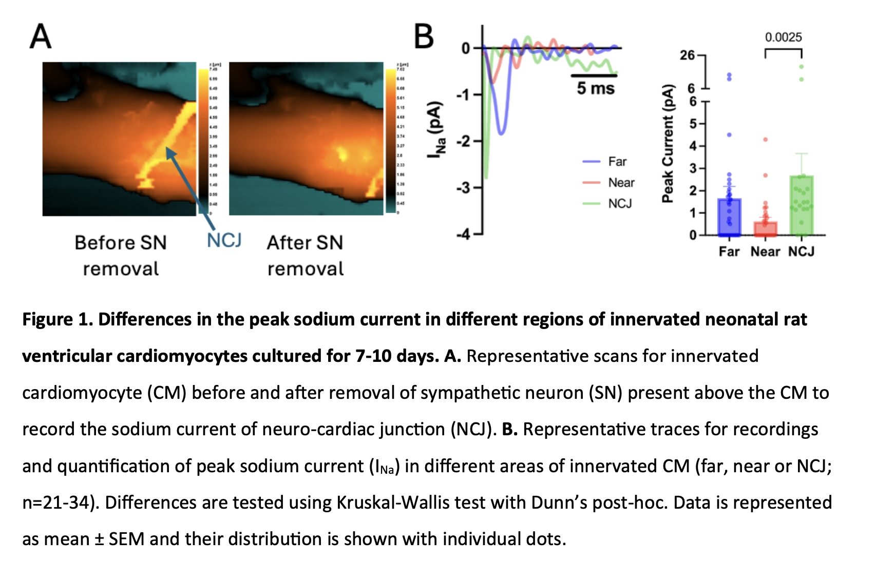
Results: The results showed that in the first 2-5 days of culture, Nav1.5 is evenly spread throughout the different regions of innervated CMs (n=7-22). Prolonged innervation with 7-10 days culture promoted redistribution of Nav1.5 in CM membrane with a preference in the NCJ region of (2.7±1.0pA) versus the lack of Nav1.5 in areas near the NCJ region (0.6±0.2pA; p=0.0025). Immunostaining also showed a similar distribution of Nav1.5 and AnkG, with the highest intensity being found in the NCJ region and the least near the NCJ region (N=3-4 replicates; n=14-16 cells). The colocalisation showed a positive correlation for the distribution of Nav1.5 and AnkG traces in both 2-5 days (r=0.7217, p<0.0001) and 7-10 days (r=0.5372, p<0.0001).
Conclusion: Collectively, the results show that innervation promotes the redistribution of Nav1.5 in innervated CM membranes, possibly by an AnkG-dependent mechanism.