Articular cartilage is avascular and exchange of nutrients and metabolic products between cells and the vasculature occurs by diffusion across distances of up to 3 mm. Studies of chondrocyte metabolism in cartilage explants show a combination of rapid aerobic lactate synthesis with a low oxygen consumption, indicating that ATP is generated predominantly by substrate-level phosphorylation in the glycolytic pathway (Lee & Urban, 1997). The supply of glucose is therefore essential if adequate levels of ATP are to be maintained for chondrocyte function. However, aside from one recent report of GLUT1 and GLUT3 isoforms of the glucose carrier in human cells (Neama et al. 2001), there have been no studies characterising mechanisms of glucose uptake by chondrocytes. In the present study, we have measured glucose transport into isolated bovine articular chondrocytes using radiolabelled deoxyglucose (DOG).
Bovine articular chondrocytes were isolated from metacarpophalangeal cartilage obtained at abattoir, using standard enzymatic digestion (Wilkins & Hall, 1995). Glucose transport was assessed by 3H-DOG uptake (1 µCi ml-1 in Hepes-buffered saline, HBS, at 37 °C) into chondrocytes (1 X 10⁶ cells ml-1) for periods of up to 30 min. Glucose influx was stopped by centrifugation (10 000 rev min-1, 10 s) and washing in ice-cold HBS. Uptake was calculated by scintillation counting and standard flux equations.
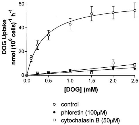
DOG uptake into chondrocytes proceeded rapidly and was a linear function of time for the first 150 s. Uptake was found to be a saturable function of extracellular DOG concentration (Fig. 1); for cells suspended in solutions supplemented with phloretin (100 µM) or cytochalasin B (50 µM), uptake was almost entirely inhibited, with only a small, linear fraction remaining. Simple fits of the phloretin-sensitive flux yielded a Km value of approximately 1 mM and a Vmax of 58 nmol (106 cells)-1 h-1. Subsequent experiments characterised the dependency on Na+ ions of glucose transport using solutions containing 2.5 mM DOG. The rate of uptake in Na+-containing solutions (54.29 ± 3.18 nmol (106 cells)-1 h-1, mean ± S.E.M., n = 3) was not significantly different (P > 0.05, Student’s unpaired t test) from that in media in which Na+ ions were replaced with NMDG (49.88 ± 2.72 nmol (106 cells)-1 h-1, n = 3).
This first report of the kinetics of glucose uptake in chondrocytes shows a system with relatively high affinity, in keeping with the circumstances of the chondrocyte, where glucose is a relatively scarce resource in the extracellular matrix. The lack of Na+ dependence and inhibition by phloretin is indicative of uptake by the glucose carrier family GLUT, and rules out a role for SGLT. Of the two isoforms of the glucose carrier demonstrated in human chondrocytes, the Km value reported here more closely fits that of GLUT3, an isoform also found to be dominant in cells from the avascular tissue of human neuronal parenchyma (Burant & Bell, 1992).
This work was supported by the Arthritis Research Campaign, UK.