ERD analysis is a tool for studying movement-related power changes in surface EEG signals (Pfurtscheller & Lopes da Silva, 1999). To study the movement-related power changes in the human basal ganglia, after ethical committee approval we assessed ERD in EEG signals from electrodes implanted in the internal segment of the globus pallidus (GPi) and in the subthalamic nucleus (STN) for deep-brain stimulation therapy in a patient with Parkinson’s disease.
The precise electrode placement was checked by MRI scans. The motor task consisted of a brisk (0.5-0.6 s) voluntary second finger extension (inter-trial interval > 10 s). Four channels were recorded for each side: surface bipolar EEG over the primary sensorimotor cortex (SMC); EEG from the GPi; EEG from the STN; and surface EMG from the contralateral extensor indicis muscle. Signals were amplified, digitized, sampled and stored on a PC for further off-line analysis. The ERD was calculated by an adaptive autoregressive identification approach (‘whale forgetting’) (Bianchi et al. 1997), through specifically developed algorithms implemented in the Matlab software. Because preliminary analysis showed that the main spectral content of the signals arising from the GPi and the STN was within 50 Hz, the ERD analysis was limited to frequencies < 50 Hz.
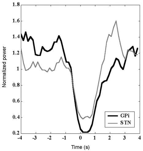
The SMC had ERDs for the 10 Hz (alpha) and the 17 Hz (low beta) frequencies, starting 0.5-0.8 s before the movement onset, peaking during the movement, recovering and progressively resuming the baseline after movement. The GPi had no ERD in the 10 Hz frequency but there was a marked ERD at 17 Hz and at 27 Hz (high beta). The STN showed ERD only in the 27 Hz frequency. Movement-related ERDs in the SMC, GPi and STN had similar time courses (Fig. 1).
In conclusion, the GPi shares the ERD at 17 Hz with the SMC, and the ERD at 27 Hz with the STN (Fig. 1). Hence the 17 Hz ERD in the SMC and GPi might functionally reflect activity in the direct pathway. Conversely the 27 Hz ERD in the STN and GPi might specifically reflect the activity in the indirect pathway of the human basal ganglia motor circuit.
This work was supported by IRCCS Ospedale Maggiore di Milano, Italy.