Cell swelling, which occurs during episodes of ischaemia and reperfusion in the heart, can be mimicked experimentally by exposing myocytes to hypo-osmotic solution. We are interested in the mechanisms which underlie cell swelling. In neonatal chick cardiac myocytes, swelling is potentiated by disruption of the actin cytoskeleton (Zhang et al. 1997), and in adult rabbit ventricular myocytes, swelling is reduced by pharmacological blockade of cationic stretch-activated channels (SACs) (Suleymanian et al. 1995), which may be located within the t-tubules (Zeng et al. 2000). In this study we have investigated the involvement of the actin cytoskeleton and SACs in the volume response of adult rat ventricular myocytes by using the actin disruptor cytochalasin-D (cyto-D) and by removing t-tubules.
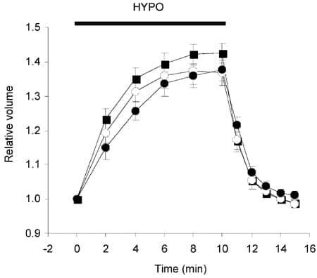
Adult Wistar rats were killed humanely and ventricular myocytes isolated enzymatically. Cells were incubated in either 10 µM cyto-D or vehicle solution (dimethyl sulphoxide 0.1 % v/v) for 1 h prior to swelling. Some myocytes were de-t-tubulated (d-Tub) (Brette et al. 2002). Cells were superfused with physiological Hepes-based solution (284 ± 1 mosmol l-1, mean ± S.E.M., n = 3) and then swollen by a 10 min exposure to a hypo-osmotic version of this solution (191 ± 1 mosmol l-1) at 22-24 °C. Cell volume was calculated from a video image of the cell.
The mean maximal increase in cell volume of cyto-D-treated or d-Tub cells was not significantly different from that in vehicle-treated cells (P > 0.05, one-way ANOVA, see Fig. 1). However, the time constant of the swelling response was significantly increased in d-Tub cells (4.21 ± 0.48 min), compared to vehicle (2.58 ± 0.09 min) or cyto-D-treated cells (2.62 ± 0.3 min) (P < 0.05 ANOVA with post hoc analysis by Tukey’s test).
We conclude that, in contrast to neonatal myocytes, the swelling response of adult cardiac myocytes is not modulated by the actin cytoskeleton. In addition, our findings suggest that the magnitude of the swelling response is not dependent upon ion channels specifically located within the t-tubules but that the absence of t-tubules slows the swelling response.
This study was funded by the BHF and the University of Leeds.