Exercise can have both a positive and negative effect on the immune system. Previous studies have shown that exercise can induce changes in many cytokines and signal molecules but the mechanism of action for these changes is not clear. Toll-like receptors (TLRs) play a crucial role in the detection of microbial pathogens by recognising products of microbial metabolism such as lipoproteins (TLRs 1, 2 and 6), zymosan (TLR2), lipopolysaccharide (LPS)(TLR4), and unmethylated CpG DNA motifs (TLR9) (Medzhitov, 2001). Following recognition of their specific ligand, TLRs expressed by antigen-presenting cells regulate the production of several cytokines including interleukin (IL)-6, IL-8 and tumour necrosis factor (TNF)-α, as well as the expression of accessory signal molecules CD80, CD86 and IL-12, that are required for the activation of naive T lymphocytes and the subsequent induction of the immune response. The effect of exercise or other forms of stress on TLRs is not well characterised.
The purpose of the present study was to examine the effects of exercise on the regulation of TLR expression and function. With local Ethics committee approval, venous blood samples were obtained from 11 healthy trained male cyclists (age 25 ± 1 years; body mass 74 ± 2 kg; maximal oxygen uptake, Î{special}J{special}max 4.7 ± 0.2 l min-1; mean ± S.E.M.) at rest, immediately after, and following 2 h of resting recovery from 90 min cycling at 65 % Î{special}J{special}max in the heat (34°C). The expression of TLRs 1, 2, 4 and 9 on monocytes was assessed by flow cytometry. Monocyte intracellular cytokine (TNF-α and IL-6) production, the expression of CD80, CD86 and MHCII was also assessed following stimulation with LPS, zymosan and poly (I:C). Data were analysed using one-way ANOVA and post hoc Tukey tests where appropriate.
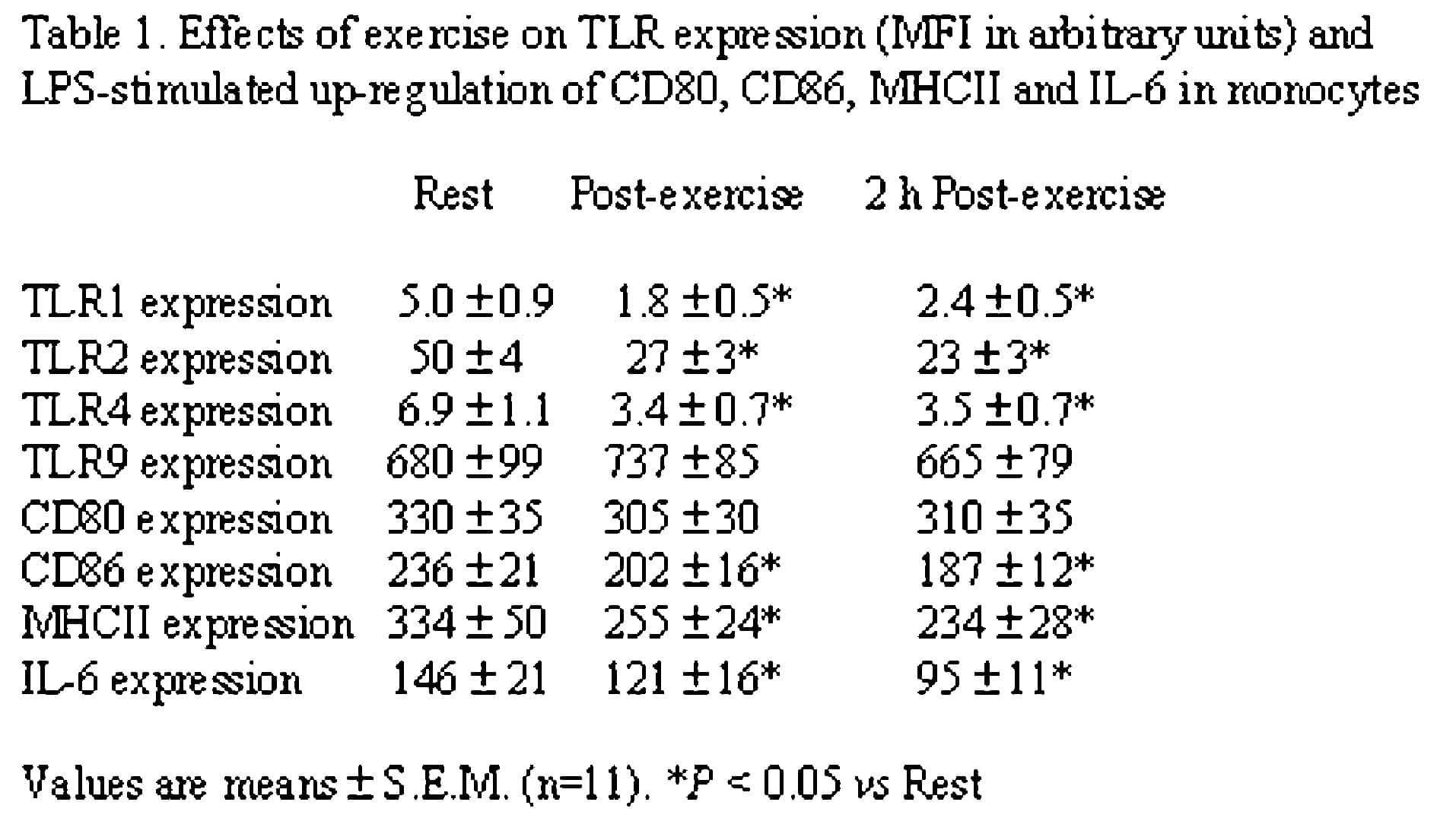
Following exercise, monocyte expression of TLRs 1, 2 and 4 (but not TLR9) was substantially decreased (P < 0.05) with little or no recovery by 2 h post-exercise (Table 1). Furthermore, the LPS-stimulated induction of CD86 and MHCII expression on monocytes was significantly lower in samples obtained following exercise compared with pre-exercise (P < 0.05). Similar results were obtained with zymosan and poly (I:C). LPS-stimulated monocyte IL-6 production was significantly reduced after exercise (P < 0.05).
These results indicate that monocyte TLR expression is reduced following an acute bout of prolonged exercise and that this is associated with decreased induction of co-stimulatory molecules and cytokines following stimulation with TLR ligands. These effects may represent a mechanism through which exercise stress impairs immune function and increases susceptibility to infection.
This work was supported by GlaxoSmithKline