A high frequency excitatory network in motor cortex generates multiple corticospinal I waves at ~ 600 Hz to a transient cortical stimulus; does the 600 Hz rhythm reflect a possible clock function present even during voluntary activity (Amassian and Stewart, 2003)? Neither individual corticospinal nor α motoneurons discharge at I wave periodicity during voluntary activity; however, a collective of discharging motor system neurons responding stochastically at the basic clock period might reveal I wave periodicity.
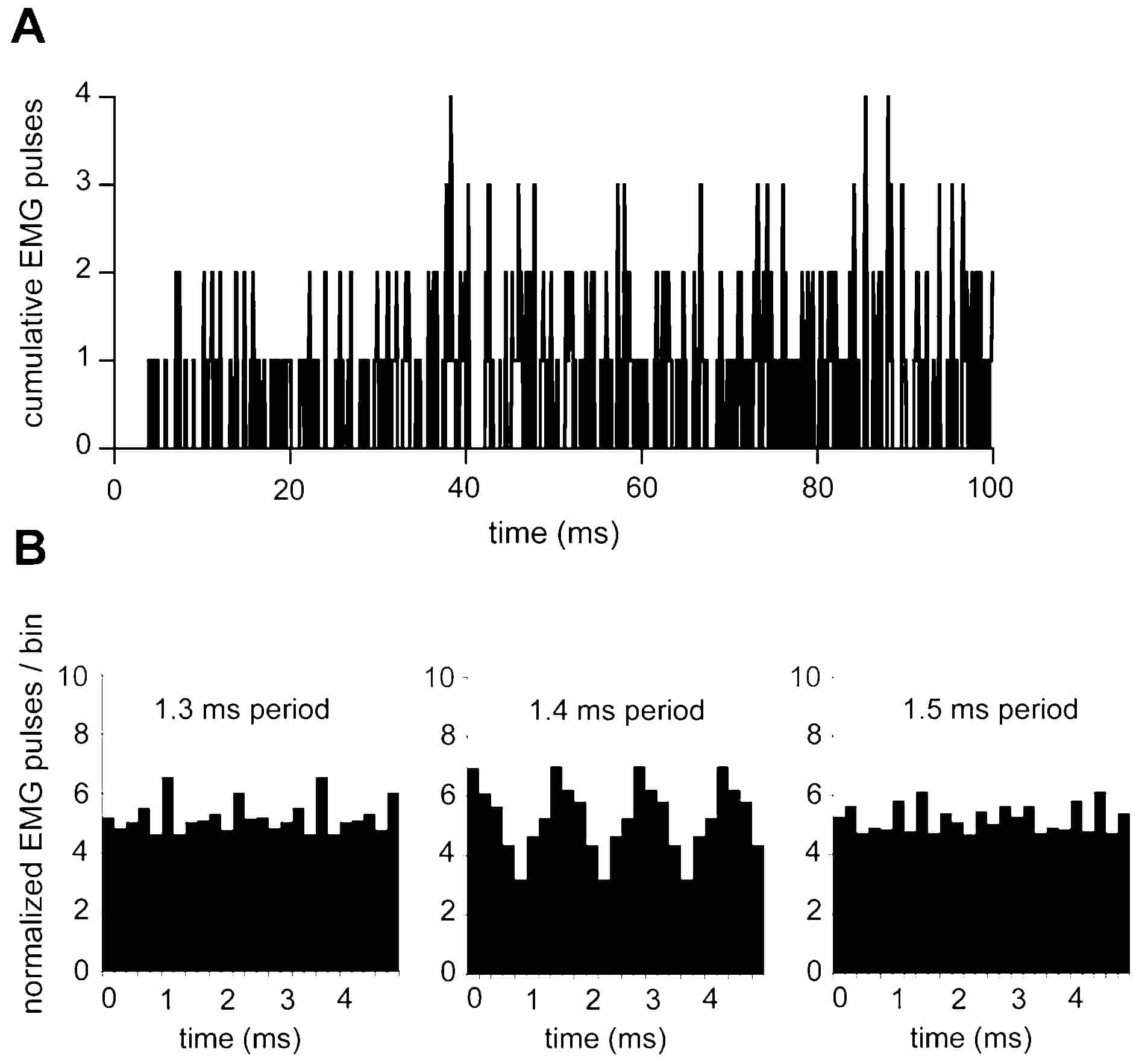
With Institutional Review Board approval and informed consent from four subjects (41-78 yrs of age), this ‘clock’ hypothesis was tested as follows: surface EMGs were recorded over sternohyoid-thyroid muscles by two silver balls, 1 mm in diameter and 10-15 mm interpolar distance; the overlying skin was cleaned with acetone and then covered with a thin layer of electrode paste. Activity by several motor units was elicited by silently articulating (without expiration) plosive consonants (e.g. D, T) and analysing the initial 100-200 ms period of activity. The EMG was half-wave rectified and converted to standard pulses; the onset of activity triggered the accumulation of a post-stimulus histogram (Fig. 1A). A dead time of 4.0 ms was introduced before the standard pulse (0.1 or 0.2 ms duration); thus intervals less than the dead time could appear only in the summed histogram.
First, the iterative expectation density (ED) function was computed on the summed histogram to identify any I wave periodicity. Iteration required that given 2 or more spike pulses in a bin, each was in turn translated to the origin before translating the contents of the next bin. Secondly, the cross-channel ED function was computed between a pulse train with a fixed period of, e.g. 1.4 or 1.5 ms and the EMG derived pulses. Pulse trains were started at the EMG onset. Cross-channel EDs were periodic for each subject at a particular pulse train period and much less periodic at neighboring periods. Figure 1B illustrates a cross-channel ED that was optimal with a pulse train period of 1.4 ms. In other recordings, the optimal period was 1.5 or 1.6 ms. In the ‘best-fit’ EDs for all subjects, the mean number of EMG pulses per bin outnumbered the in-phase increment.
Summarizing, during articulation, the discharges of at least some EMG units are related to I wave like periodicity.