
Changes observed in skeletal muscle metabolism during sepsis are consistent with impaired oxidative metabolism, and in particular to a defect in pyruvate utilisation, due possibly to an inhibition of the pyruvate dehydrogenase complex (PDC; Vary et al. 1999). Phosphorylation (inactivation) of the PDC is regulated by pyruvate dehydrogenase kinase (PDK), of which isoforms 2 and 4 are the most prevalent in skeletal muscle. The aim of the present study was to examine the effect of lipopolysaccharide (LPS) infusion on PDK2 and PDK4 mRNA expression in fast- and slow-twitch skeletal muscle of the rat. Male Sprague-Dawley rats (previously implanted with jugular venous catheters under general anaesthesia (fentanyl and medetomidine, 300 μg kg-1 each i.p.)), were divided into 6 groups, and infused with either saline (0.4 ml h-1) for 2 h (n=8), 6 h (n=7) or 24 h (n=8), or LPS (E. coli serotype 0127:B8, dissolved in saline, 150 μg kg-1 h-1) for 2 h (n=8), 6 h (n=8) or 24 h (n=6). At each time point, the slow-twitch soleus and fast-twitch extensor digitorum longus (EDL) muscles were freeze clamped in situ under terminal anaesthesia (sodium pentobarbital, i.v.) and stored at -80°C. All animal procedures complied with Home Office regulations. Total RNA was extracted from muscle samples and expression of PDK2 and PDK4 determined by real-time PCR. The results are presented in Table 1. LPS infusion resulted in a marked and time-dependent increase in PDK4 mRNA expression in EDL and soleus, whereas a less marked time-dependent decrease of PDK2 expression was observed in soleus (Table 1). These findings are consistent with down regulation of carbohydrate oxidation following LPS administration, which, based on the present data, may be due to PDC inactivation as a consequence of a rapid and marked increase in PDK4 mRNA and presumably protein expression.
King's College London (2005) J Physiol 565P, C99
Communications: Lipopolysaccharide infusion rapidly alters pyruvate dehydrogenase kinase mRNA expression in rat skeletal muscle
Alamdari, N ; Murton, A J; Constantin-Teodosiu, D ; Gardiner, S M; Bennett, T ; Layfield, R ; Greenhaff, P L;
1. School of Biomedical Sciences, University of Nottingham, Nottingham, United Kingdom.
View other abstracts by:

Table 1: Fold changes in PDK2 and PDK4 mRNA expression from the corresponding control value in EDL and soleus muscles of the rat after LPS infusionValues are mean ± S.E. ∗ Significantly different from 2h LPS treated muscle (P<0.05 MANOVA LSD post-hoc). † Significantly different from 6h LPS treated muscle (P<0.05 MANOVA LSD post-hoc). ‡ Significantly different from saline control at corresponding time point (P<0.05 MANOVA).
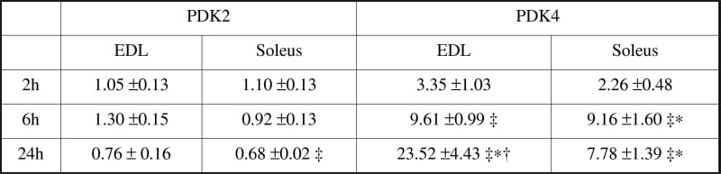
Table 1: Fold changes in PDK2 and PDK4 mRNA expression from the corresponding control value in EDL and soleus muscles of the rat after LPS infusionValues are mean ± S.E. ∗ Significantly different from 2h LPS treated muscle (P<0.05 MANOVA LSD post-hoc). † Significantly different from 6h LPS treated muscle (P<0.05 MANOVA LSD post-hoc). ‡ Significantly different from saline control at corresponding time point (P<0.05 MANOVA).
Where applicable, experiments conform with Society ethical requirements.