
Time of day increments in strength have previously been reported (Deschenes et al. 1998; Martin et al. 1999), but no attempt to study this effect in conjunction with internal muscle structure has been made, even though in vivo, muscle architecture (fibre pennation angle (θ) and muscle size), are determinants of the capacity of a muscle to generate torque (Maganaris et al. 2001). The aim of the current study was therefore to examine whether muscle architecture can account for a) force changes or b) any changes in the torque-angle relationship with time of day. Sixteen healthy young men (aged 23 ± 5.7 years) were tested at 7h45am and 5h45pm. To prevent order effects, seven subjects were tested in the order am to pm, and the rest in the order pm to am. Knee extensors test angle was randomised and the best of 3 contractions (peak isometric extension torque (PIET)) at each angle was used for analysis. The investigation was approved by the Manchester Metropolitan University Institutional Ethics Committee and all subjects gave their written informed consent to participate in the study. PIET showed an average 7.0 ± 1.8% (p=0.023) upward shift throughout the range (30-90 deg) in the evening compared to the morning. The polynomial regressions fitted through the torque/angle relationship data showed a 10 deg shift of the angle at which PIET occurs (from ~80 deg at am to ~70 deg at pm) so that PIET was seen at shorter muscle lengths in the evening. Also at the knee angle related to the PIET, in the contracted muscle a 14 ± 2.0% (p=0.002, paired t test) time-of-day related change in θ (to a greater θ at pm compared with am) was observed. Muscle architecture was measured both at rest and at a standardised force level (250 N), both am and pm. In the resting muscle, increments of 8 ± 1.2% (p=0.001) in muscle thickness, and 13 ± 5.1% (p=0.046) in pennation angle (see Fig. 1), were observed at pm relative to am. In a subset (n = 5) a trend for a decrement in the pennation angle (- 5.6%) was observed from am to pm during the standardised exertions. These results thus suggest that the time-of-day differences in muscle extensor torque are partly explained by changes in muscle architecture.
King's College London (2005) J Physiol 565P, PC27
Communications: Human knee-extensors architecture: diurnal rhythmicity and torque characteristics
Graham - Smith, Philip ; Pearson, Stephen John; Bruce, Stuart A; Onambele, Gladys ;
1. Directorate of Sport, University of Salford, Manchester, United Kingdom. 2. School of Biomedical Sciences, Kings College, London, United Kingdom. 3. Department of Exercise & Sport Science, Manchester Metropolitan University, Manchester, United Kingdom.
View other abstracts by:
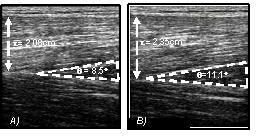
Figure 1. Typical sonographs of the sagittal plane of the vastus lateralis muscle at rest with the knee at an angle of 85Deg (right angle is 90Deg). A) AM and B) PM showing pennation angle (theta) and muscle thickness (t).
Where applicable, experiments conform with Society ethical requirements.