
Cardiomyopathy and arrhythmias are major causes of mortality in iron poisoning and chronic iron overload (Cohen et al. 2004). However, direct evidence on how toxic concentrations of iron modify cardiac cellular function is scarce. We have examined the acute effects of a large dose of iron (as Fe2+) on Ca2+ handling in isolated rat ventricular myocytes. Rats were humanely killed. Single cardiac ventricular myocytes were isolated by collagenase/protease digestion and loaded with the Ca2+-sensitive fluorescent dye fluo-3. Cells were field-stimulated at 0.33 Hz and cell shortening and Ca2+ transients were recorded. Prolonged exposure (30 min) of cells to 500 μM iron (II) (added as iron (III) chloride in the presence of 1 mM of the reducing agent ascorbate) led to a pronounced increase in both the systolic Ca2+ transient amplitude and cell shortening (Fig. 1). This was followed after a few minutes (16 ± 8 min, mean ± S.E.M., n = 10) by the appearance of spontaneous Ca2+ waves (and propagating waves of contraction), which increased in frequency leading to cell contracture and death. Preloading the cells with the iron chelator deferiprone (DFP; 100 μM) attenuated, but did not completely prevent, the effects of iron (mean time to onset of spontaneous activity in DFP 21 ± 6 min, n = 7, n.s. vs. iron, p>0.3 by t test; frequency of spontaneous contractions 6.5 ± 1.4 min-1 in iron, 3.1 ± 2.4 min-1 with DFP, significantly different with p<0.03). In non-stimulated (quiescent) cells, iron treatment led, again after a delay, to the onset of spontaneous contractile activity. Our results show that iron (II) has acute effects on ventricular cell Ca2+ handling that could be arryhthmogenic. The mechanism of these effects is unknown but may be related to the action of oxidative radicals on Ca2+ transport processes at the sarcoplasmic reticulum or the sarcolemma.
University of Bristol (2005) J Physiol 567P, C30
Oral Communications: Acute exposure to iron (II) alters Ca2+ handling in isolated rat ventricular myocytes
Baptista-Hon, Daniel T.; Elliott, Austin Charles; Diaz, Mary E.;
1. Univ of Manchester, Manchester, United Kingdom. 2. Veterinary Biomedical Sciences, Edinburgh University, Edinburgh, United Kingdom.
View other abstracts by:
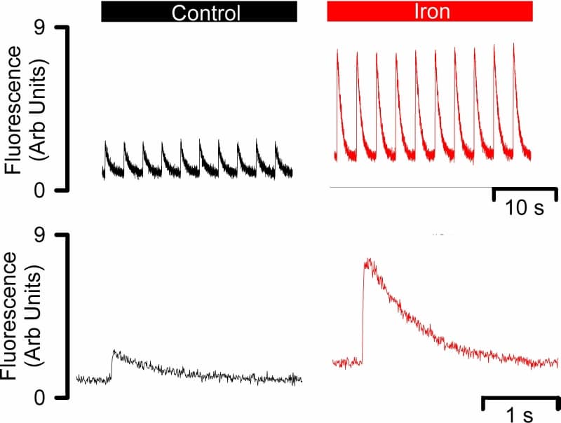
Figure 1. Effects of acute iron exposure on the systolic Ca transient. Original data show: top systolic Ca transients in response to electrical stimulation every 0.33 Hz in control (left) and iron (right). Bottom average systolic Ca transients taken from the data shown above in control (left) and in iron (right).
Where applicable, experiments conform with Society ethical requirements.