
The process of mechanotransduction is essential for the health and homeostasis of the articular cartilage. However, the underlying signalling pathways are poorly understood. This study examines the influence of cyclic mechanical compression on chondrocyte Ca2+ signalling and the involvement of mechanosensitive release of cellular ATP. The study used a well-characterised model consisting of isolated bovine (from an abattoir) articular chondrocytes cultured in agarose constructs [1]. After 24h, constructs were incubated in lluo-4 AM (Molecular Probes) and mounted in a custom made compression rig [1] on a confocal microscope (Perkin Elmer). Cyclic compression at 1Hz between 0% and 10% strain was applied for 1, 10, 100 or 300 cycles. This was followed by a 5min period in which images of fluo-4-labelled cells, approximately 20 cells per field of view, were captured every 4s using a ×20 objective. Separate constructs were held at 10% static compression and subjected to an identical 5min imaging period in the compressed state. Control constructs remained unstrained. In further studies constructs were bathed in apyrase (10 units/ml) during 1Hz cyclic compression for 10 cycles and throughout the subsequent 5min imaging period. Unstrained controls were also bathed in apyrase. Student’s t tests (p<0.05) were used to compare the percentage of cells showing Ca2+ transients in samples of individual constructs (n=8-15). In unstrained agarose constructs, 50% of cells exhibited spontaneous Ca2+ transients over the 5min period. Static 10% compression produced no change in Ca2+ signalling. Previous studies have shown increased signalling at 20% compression [1] indicating that the effect may be strain magnitude dependent. Cyclic compression for 1, 10 or 100 cycles was followed by a statistically significant increase in the percentage of cells exhibiting Ca2+ transients (Fig. 1). However, 10 or 100 cycles did not induce additional responses compared to 1 cycle, suggesting a redundancy of signal transduction. The involvement of a paracrine/autocrine mechanism was confirmed by the finding that the ATP scavenger, apyrase, abolished the up regulation of Ca2+ signalling produced by cyclic compression. After 300 cycles, Ca2+ signalling was not significantly different to that in unstrained controls, possibly associated with ATP receptor fatigue. In conclusion, cyclic compression activates Ca2+ signalling via mechanosensitive release of ATP as part of a chondrocyte mechanotransduction pathway.
University of Bristol (2005) J Physiol 567P, C52
Oral Communications: Mechanical compression activates chondrocyte calcium signalling in a cycle-dependent manner involving the release of ATP
Knight, Martin Matthew; Pingguan, Belinda; Lee, David; Bader, Dan;
1. Medical Engineering Division, Dept of Engineering, Queen Mary, University of London, London, United Kingdom.
View other abstracts by:
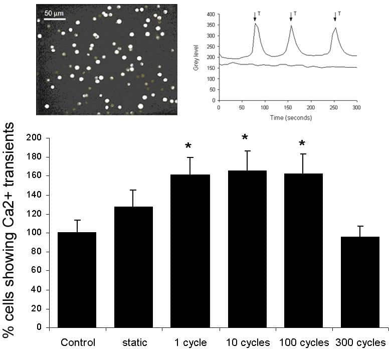
Figure 1. Percentage of cells exhibiting calcium transients in 5min period during static compression or following cyclic compression for 1 10 100 or 300 cycles. Values represent mean ± S.E.M. normalised to unstrained control (*p<0.05). Inset shows image of fluo-4-labelled cells and represenative Ca2+ transients for a single cell.
Where applicable, experiments conform with Society ethical requirements.