
Voltage-dependent calcium channels (VDCC) are multi protein complexes consisting of at least three subunits (α1, β, α2 δ). VDCC β subunits are cytosolic proteins involved in surface expression of α1 subunit. Co-expression of β subunits increase current amplitude manifold due to the increase in availability of α1 subunits at the membrane (Brice et al. 1997). In addition, β subunits have modulatory effects on channel properties, i.e. they hyperpolarize the voltage dependence of activation and differentially affect inactivation kinetics of the channel (Olcese et al. 1996). VDCC play an important role in neurotransmitter release, gene expression and excitation-contraction coupling. Therefore, they are attractive targets for treating disorders associated with these functions. In recent history, the search for calcium channel blockers has centred around the α subunit. However, due to poor selectivity, many such compounds have not succeeded in development. Hence, the requirement is for new or next-generation calcium channel modulators or inhibitors. Here, we report the development of an assay to identify a novel class of compounds that inhibit protein-protein interactions between α1 subunit and the β subunit that can be used to treat diseases associated with smooth muscle constriction, such as urinary incontinence, hypertension and angina. The full-length Cav β3 subunit and the α interaction domains (AID) of Cav 1.2 and Cav 2.2 were amplified from rat brain cDNA. Cav β3 subunits were subsequently expressed in E. coli and immobilised onto streptavidin substrates via the affinity tag, biotin carboxyl carrier protein. The purified AID proteins were Cy3-labelled and used in binding experiments to demonstrate assay principle. Cav β3 protein was successfully immobilised on streptavidin-coated plates. Furthermore, an anti-Cav β3 antibody directed towards the C-terminal epitope demonstrated that the immobilised Cav β3 subunits were full-length. Increasing concentrations of Cy3-Cav 1.2 or 2.2 AID proteins were incubated with Cav β3, a saturable binding was observed with good signal to noise ratio. In order to define the assay principle, binding affinity and kinetics between Cav AIDs and Cav β3 subunits were characterised further (Table 1). In summary, an assay has been developed that allows the determination of the affinity and binding kinetics of the interaction between Cav1.2 and Cav2.2 AIDs to the full-length immobilised Cav β3 subunit to be studied in a micro-well format. Utilising this assay for high-throughput screening has the potential to identify compounds that modulate VDCCs through a novel mode of action.
University of Oxford (2005) J Physiol 568P, PC16
Poster Communications: In vitro assay for identifying modulators of Cav channel α1–β subunit interactions
Kanumilli, Srinivasan; Ginham, Rachel; Stafford, Simon; Hogg, Dayle; Kozlowski, Roland;
1. Pharmacology, University of Bristol, Bristol, United Kingdom. 2. Lectus Therapeutics Ltd, Bristol, United Kingdom.
View other abstracts by:
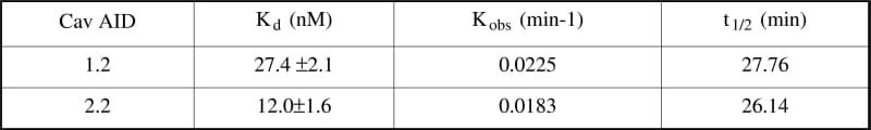
Table 1. Binding parameters of Cav 1.x AID domains to immobilised Cav β3 proteins. Kd equilibrium dissociation constant (means ± S.E.M. Kobs observed association rate constant; t1/2 amount of time required for the ligand to occupy 50% of the binding sites. n=3.
Where applicable, experiments conform with Society ethical requirements.