
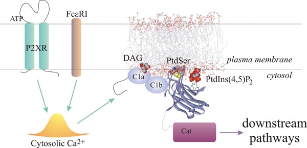
C2 domains are conserved protein modules widely distributed in many eukaryotic signaling proteins being PKCs among them (1, 2). The C2 domains of classical PKCs bind to membranes in a Ca2+-dependent manner and thereby act as cellular Ca2+ effectors. Recent findings suggest that the C2 domain of PKCα interacts specifically with PtdIns(4,5)P2, through its lysine rich cluster, with a higher affinity than that obtained with POPS-containing vesicles (3, 4). In this work, we performed a comparative study between the three C2 domains of classical PKCs. Isothermal titration calorimetry has revealed that the C2 domains of PKCα and β display a double affinity to bind to PtdIns(4,5)P2-containing vesicles than the C2 domain of PKCγ. Comparative studies by using lipid vesicles composed of both POPS and PtdIns(4,5)P2 as ligands revealed that the domains behave as PtdIns(4,5)P2-binding modules rather than POPS-binding modules, demonstrating that the presence of the phosphoinositide in membranes increases the affinity of the domain about 3.5-fold in PKCγ, 4.5-fold in PKCβ and 9.5-fold in PKCα. When their Ca2+-dependences for membrane binding were analyzed it was observed that in the presence of PtdIns(4,5)P2 all C2 domains decreased their Ca2+ needs, although the most significant was PKCβ with a 36-fold decrease compared to 8.5- and 5-fold decreases exhibited by PKCγ and PKCα, respectively. In vivo experiments using differentiated PC12 cells transfected with each C2 domain fused to ECFP and stimulated with ATP demonstrated that at limiting intracellular [Ca2+], the three C2 domains translocate to the plasma membrane at very similar rates, but however, the plasma membrane dissociation event is different among them, being PKCα the isoenzyme that persists for a longer time in the plasma membrane, reflecting their different Ca2+ needs and affinities for PtdIns(4,5)P2.
Life Sciences 2007 (2007) Proc Life Sciences, C80
Research Symposium: The C2 domains of classical PKCs are specific PtdIns(4,5)P2-sensing domains.
S. Corbalan1, M. Guerrero-Valero1, C. Marin-Vicente1, J. C. Gomez-Fernandez1
1. Bioquimica y Biologia Molecular A, Universidad de Murcia, Murcia, Spain.
View other abstracts by:
Where applicable, experiments conform with Society ethical requirements.