
Interleukin-1 (IL-1) is a pro-inflammatory cytokine that exacerbates various forms of neurodegeneration, including alpha-amino-3-hydroxy-5-methyl-4-isoxazolepropionate (AMPA) induced excitotoxic neuronal death. While injection of AMPA alone into the rat striatum results in localised neuronal loss in the striatum, co-injection of AMPA plus IL-1 (A+IL-1) causes extensive neuronal loss in both the striatum and cortex. This is despite the fact that striatal injection of IL-1 alone produces no perceptible neuronal loss 1. Injection of IL-1 into the brain is known to induce neutrophil recruitment2, and several studies suggest that neutrophils contribute to neuronal loss following stroke3,4. The aim of this study was therefore to test the hypothesis that neutrophils contribute to IL-1-mediated exacerbation of AMPA-induced excitotoxic injury. Neutrophil depletion serum, or normal rabbit serum as a control, was injected into the lateral tail vein of male Sprague-Dawley rats 24h and 1h prior to intra-striatal injection of A+IL-1 (under 3% isoflurane anaesthesia in a mixture of 1.2l/min nitrous oxide and 0.6l/min oxygen; anaesthesia was then maintained by 1.5-2.5% isoflurane in the same nitrous oxide/oxygen mix throughout the procedure). The animals were then sacrificed at 4, 8 or 24h after injection of A+IL-1 and the brain and serum removed. Immunohistochemistry was used to detect neutrophils and c-fos protein expression (to denote neuronal activation) within the parenchyma. Fluoro-jade B was used to detect degenerating neurons at 4 and 8h post-injection. In animals sacrificed at 24h, brain lesion volume was calculated from cresyl violet stained coronal sections. The most noteworthy finding was that neutrophil depletion prior to intra-striatal injection of A+IL-1 significantly reduced the size of the resultant lesion. In the cortex the lesion was reduced by 68.3% and sub-cortical damage was reduced by 40.5% which gave a 60% smaller total lesion volume in neutrophil depleted animals compared to non-neutrophil depleted control animals (Fig 1A). Typically, the cortical neurons salvaged by prior neutrophil depletion were in the deeper cortical layers (Fig 1B). Our findings regarding neutrophil recruitment and neuronal activation and damage at earlier time-points will be presented.
Life Sciences 2007 (2007) Proc Life Sciences, PC122
Poster Communications: Interleukin-1 exacerbates excitotoxic brain injury via a neutrophil-dependent mechanism
L. M. Mccluskey1, S. M. Allan1
1. FLS, University of Manchester, Manchester, United Kingdom.
View other abstracts by:
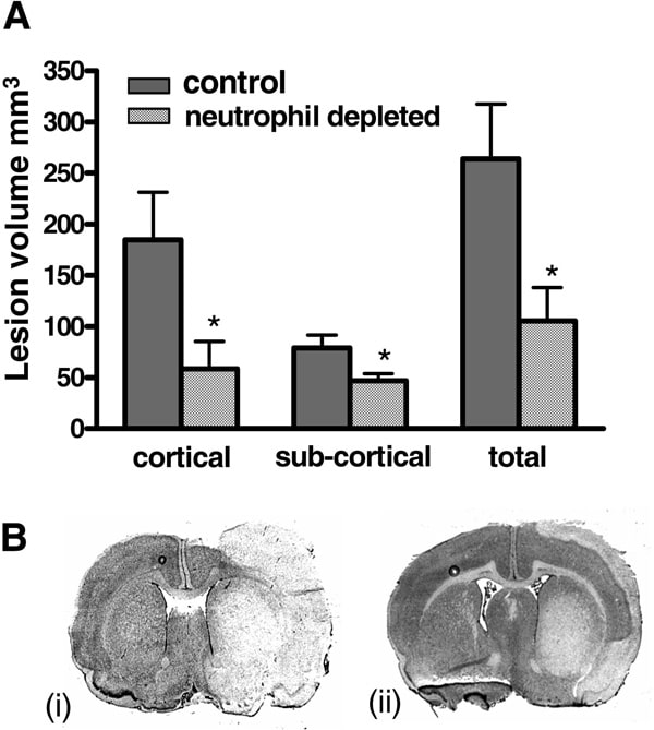
Fig 1. Effect of neutrophil depletion on A+IL-1 induced lesion. (A) A+IL-1 induced lesion volume *P<0.05 Students t test. (B) Cresyl violet stained 20μm coronal sections showing typical A+IL-1 induced lesion (area of pallor) in striatum and cortex of non-neutrophil depleted (i) and neutrophil depleted (ii) animals
Where applicable, experiments conform with Society ethical requirements.