
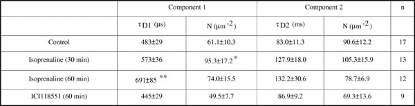
The β3-adrenergic receptor (β3-AR) is one of three G-protein coupled receptors which mediate the physiological effects of the catecholamines adrenaline and noradrenaline. In common with the β1– and β2-adrenoceptors, the β3-AR mediates its effects through activation of stimulatory (Gs) G-proteins to increase cellular levels of cAMP (Arch, 2001). However, unlike these closely related receptors, the β3-AR is resistant to short term desensitisation and internalisation following agonist exposure (Strosberg, 1997). Here, we have investigated the effect of agonist exposure on the diffusion of the β3-AR in the membrane microdomains of individual cells using fluorescence correlation spectroscopy (FCS). The β3-AR was fused via its C-terminus to eGFP in the vector pcDNA3.1. This vector was transfected transiently into Chinese hamster ovary cells (CHO-β3GFP cells) using Lipofectamine, according to manufacturer’s instructions. Cells were exposed to the β3-AR agonist isoprenaline (10-5M) or the antagonist ICI118551 (10-5M) at 37°C as indicated, prior to FCS measurements being taken on the upper cell membrane, as described previously (Briddon et al., 2004). Measurements were taken for 2x15s following a 15s pre-bleach, and autocorrelation data anlysed in Zeiss AIM software. Autocorrelation curves from FCS measurements on the upper cell membrane of CHO-β3GFP cells were best fit assuming two diffusion components (τD1 = 396±17μs and τD2 = 82.6±6.4ms, n=52). Of the total diffusing receptor population, approximately 60% was present as the faster-diffusing component (Particl numbers for τD1 = 37.1±4.6 and for τD2 = 53.0 ± 6.4 μm-2). As shown in Table 1, exposure to isoprenaline for 60 min caused an increase in τD1, indicating a slowing of receptor diffusion. This was preceeded by an increase in the number of receptors showing that slower diffusion time. In contrast, antagonist exposure had no significant effect on receptor diffusion. These results indicate that, whilst the β3-AR may not internalise readily, agonist stimulation causes a decrease in the diffusivity of the receptor within the cell membrane. This slowing could be a result of receptor aggregation, or possibly agonist-induced movement of receptor between differing membrane compartments.
Life Sciences 2007 (2007) Proc Life Sciences, PC472
Poster Communications: Membrane diffusion of the β3-adrenoceptor is slowed by agonist stimulation
S. Briddon1, S. Hill1
1. Institute of Cell Signalling, University of Nottingham, Nottingham, United Kingdom.
View other abstracts by:
Where applicable, experiments conform with Society ethical requirements.