
Within intracardiac ganglia (ICG) of adult animals different neurone types have been identified and classified according to their electrophysiological properties, the nature of the synaptic input they receive, and their morphological characteristics. These differences have been proposed to underpin differences in function. The intrinsic properties of a neurone are not static, but can alter during development by the up- or down-regulation of expression of ion channels and their subunits. Recent reports have documented developmental changes in expression of GABAA receptor channels and SK channels in rat ICG neurones (Fischer et al., 2005; Harper & Rimmer, 2006). Diversity in the electrophysiological and morphological characteristics have been observed in ICG neurones from different mammals. We have carried out studies to determine the neuronal types in the neonatal (5-9 days) and adult (> 6 weeks) BALB/c mouse. This information will be used to organize the characterization of specific genetically modified animals. The electrophysiological properties of mouse ICG neurones undergo postnatal differentiation. Comparison of the discharge activity evoked by depolarizing currents in neonates and adults reveals marked differences. Neurones of neonates (6/6) fire only a single action potential over a large range of steps, but neurones in adult ganglia exhibit a wide range of activities from phasic (7/17) , multiple adapting (8/17) to tonic discharge (2/17). The input resistance of neonates was in several instances unexpectedly similar to that of adults. Cell capacitance (Ci) was calculated from Ci = τm / Ri to provide an index of the size of the neurone. The reduction in Ri and increased Ci measured for neurones in the neonatal ganglion is consistent with gap junction coupling, increasing their apparent electrical size. Dye coupling (Neurobiotin) was found in 2/6 neurones in neonates but not in adults. These studies indicate changing patterns of expression of ion channels with maturation of the cardiac nervous system
Life Sciences 2007 (2007) Proc Life Sciences, PC531
Poster Communications: Developmental changes in the electrophysiological properties of intracardiac ganglion neurones of the mouse in situ
L. M. Wallace2, P. E. Durante1, A. A. Harper1
1. Molecular Physiology, University of Dundee, Dundee, United Kingdom. 2. Pathology and Neuroscience, University of Dundee, Dundee, United Kingdom.
View other abstracts by:
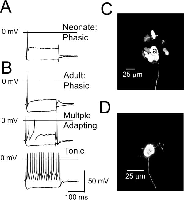
Voltage responses obtained in response to long (200 ms) de- and hyperpolarizing current pulses (2x threshold and -0.1 nA) recorded from neonatal (A) and adult ICG (B) neurones. Confocal images of a neonatal (P5) mouse intracardiac ganglion (C) showing presumptive evidence for dye-coupling between neurones (neurone ‘a’ was injected with the gap junction permeable tracer Neurobiotin which spread to neighbouring neurones) and an adult mouse intracardiac ganglion neurone (D).
Where applicable, experiments conform with Society ethical requirements.