
Cats, dogs and humans show a consistent pattern of intercostal muscle discharges in their dorsal regions, the external layer being inspiratory, the internal layer expiratory (De Troyer et al. 2005). Here we report that the pattern in the rat is different. Intercostal spaces T6-T10 were studied in vagotomised rats, stimulated with CO2 to give a strong respiratory drive. Intercostal nerve discharges were recorded in anaesthetized or decerebrate rats, under neuromuscular blockade, as were intracellular recordings from motoneurones with axons in intercostal nerves of T8 or T9. EMG recordings were made from proximal parts of spaces T6-T10 in spontaneously breathing anaesthetized animals. Anaesthetics: ketamine/xylazine, induction I.P., 100mg/kg, 10mg/kg respectively, maintenance I.V., as required, same ratio; urethane, induction 1.4 g/kg I.P., maintenance 0.2 g/kg I.P. if needed; alpha-chloralose, induction and surgery under halothane, titrated to alpha-chloralose I.V. up to 80 mg/kg for recordings. Decerebration: induction and surgery under halothane or ketamine/xylazine (as above), brain removed rostral to colliculi following other surgery. Neuromuscular blockade: pancuronium bromide I.V., initially 2.5 mg/kg, then 1.7 mg/kg.hr (adequate anaesthesia assured by stability of blood pressure, heart rate and pattern of respiratory discharges following noxious stimuli). Under ketamine/xylazine or urethane, expiratory discharges were seen only in relatively lightly anaesthetized animals (cf. Saywell et al. 2007) and were weak or intermittent. Only under alpha-chloralose or decerebration were these discharges strong and regular. In these conditions the following were observed (see Fig. 1). ● External intercostal nerve discharges showed a biphasic pattern of discharges, with an expiratory burst in addition to the inspiratory one (cf. Tian & Duffin, 1996). ● EMG recordings from the most proximal region of the internal intercostal muscle showed a predominantly inspiratory pattern, as did recordings from the internal intercostal nerve branch that innervates it, though when expiration was strong the pattern was biphasic. Denervation of neighbouring intercostal spaces, plus destruction of the external intercostal layer and of multiple levator costae muscles (inspiratory) confirmed that the EMG activity arose from the sampled area. Some motor units were active in both the expiratory and the inspiratory bursts. ● Intracellular recordings included 2/3 external intercostal nerve motoneurones with expiratory depolarizations and 19/56 internal intercostal nerve motoneurones with biphasic depolarizations. The biphasic patterns often appeared to include simultaneous excitation and inhibition during inspiration. The spatial distribution of these patterns and their functional significance have yet to be explored.
University of Leeds (2008) Proc Physiol Soc 10, C11 and PC60
Oral Communications: Patterns of respiratory activation for intercostal muscles in the rat
A. de Almeida1, P. A. Kirkwood1
1. Sobell Dept Motor Neuroscience & Movement Disorders, UCL Institute of Neurology, London, United Kingdom.
View other abstracts by:
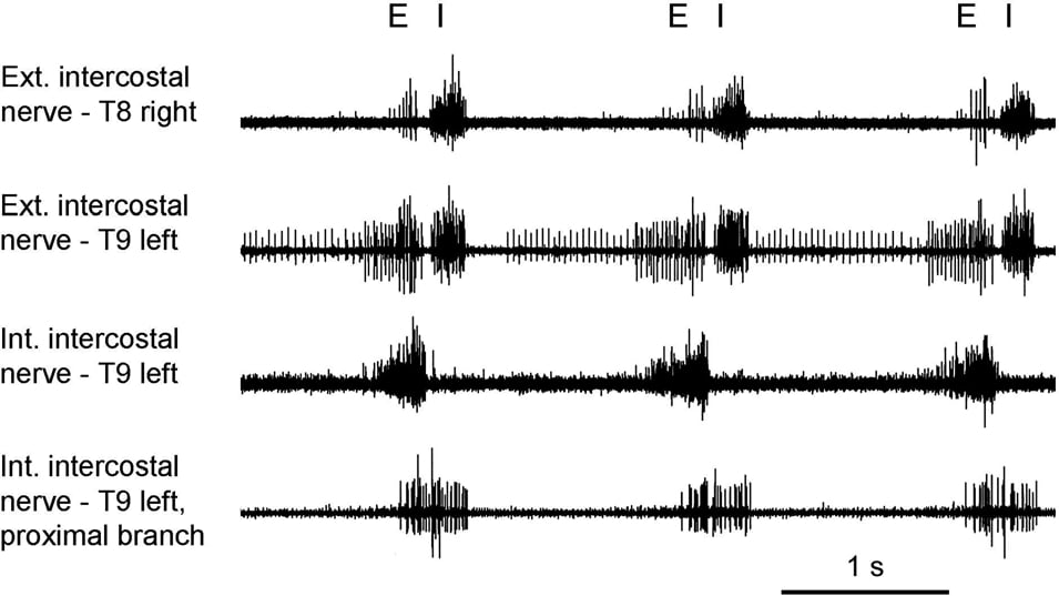
Figure 1. E, expiration; I, inspiration (chloralose). De Troyer A et al. (2005) Physiol Rev 85, 717-756.
Where applicable, experiments conform with Society ethical requirements.