
It is well-established that the neuronal plasma membrane Ca-ATPase carries H+ ions into the cell at the same time as it pumps out Ca2+ ions (Schwiening et al, 1993, see also Niggli & Sigal 2008). This results in pH changes both inside and outside cells. I have recently reported that the coupling ratio in snail neurones at pHo 7.5 is close to 1Ca:2H, in agreement with much but not all earlier work (Thomas, 2009). The PMCA Ca:H coupling ratio in erythrocytes and barnacle muscle has been reported to vary with pHo from about 1:2 at pH 6.5 to 1:0 at pH 8.5 (Milanick, 1990) or from 1:3 at pH 6.5 to 1:1 at pH 8.2 (DeSantiago et al 2007). I have now, using the same methods as previously reported, measured the effects of increasing or decreasing pHo by one unit on the coupling ratio in snail neurones. I injected Ca2+ iontophoretically and measured the resulting intracellular pH (pHi) changes at pHo values of 6.5, 7.5 and 8.5, as shown in Fig 1. In this representative experiment each injection caused a fall in pHi which was little different when the external pH was increased or decreased by one pH unit. In a total of 5 similar experiments, in some of which I also measured intracellular Ca2+ changes, I made 29 injections at the control pHo 7.5, 17 injections at pH 8.5 and 15 at pH 6.5. I found that the average fall in pHi per unit charge injected at pH 8.5 was 0.93 ± 0.06 (S.E.M., n = 5) of that for the control pH, while at pH 6.5 the average was 1.04 ± 0.06 of control. These findings are very different to those previously reported for red blood cells and barnacle muscle.
University of Manchester (2010) Proc Physiol Soc 19, C28
Oral Communications: Changing external pH has no effect on the Plasma Membrane Calcium ATPase (PMCA) coupling ratio in snail neurones.
R. C. Thomas1
1. PDN, University of Cambridge, Cambridge, United Kingdom.
View other abstracts by:
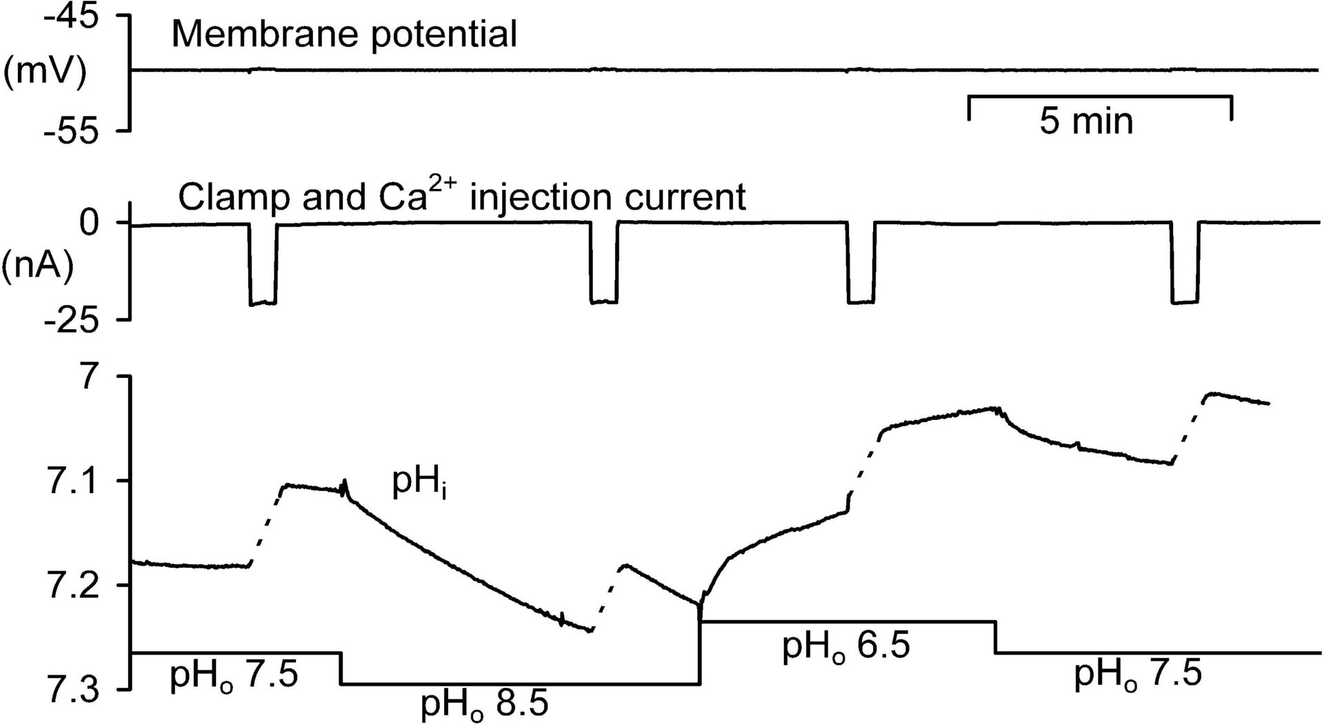
Fig 1. Experiment on a large neurone in an isolated snail ganglion bathed in normal snail Ringer buffered to pH 7.5, 8.5 and 6.5. The membrane potential (top trace) was recorded and clamped (current shown in second trace) with conventional microelectrodes filled with 1 M CsCl. Intracellular pH was recorded with a pH-sensitive microelectrode, and intracellular Ca2+ injections were made iontophoretically via a microelectrode filled with 0.1 M CaCl2. During these injections the pHi record was obscured by electrical interference, so a dashed line has been drawn from the start of each injection to a few seconds after the end.
Where applicable, experiments conform with Society ethical requirements.