
Introduction Individuals with type 2 diabetes have a risk of heart failure 2-5 times greater than age matched controls, and the risk is greater for women compared to men. Prior to the onset of heart failure, individuals with type 2 diabetes exhibit structural and functional abnormalities of the myocardium. Although exercise training improves myocardial function in animal models of type 2 diabetes, data is in humans is limited. This study aimed to investigate the effect of moderate intensity exercise training upon cardiac function in overweight and obese postmenopausal women with type 2 diabetes (T2) compared to BMI matched, healthy controls (ND). Methods Seven T2 and 8 ND women (BMI > 25 kg/m2) volunteered for the study; all were postmenopausal (mean age 57 ± 4.9 years). Prior to and following 6 months of moderate intensity exercise training (55-75% heart rate reserve), participants completed a modified Bruce cardiopulmonary exercise test to assess VO 2peak. Cardiac function (resting and peak cardiac power output; pkCPO, and cardiac reserve; C-res) were calculated from cardiac output, determined by non -invasive inert gas re-breathing, and mean arterial pressure (MAP) determined by manual sphygmomanometry. Anthropometry (body mass, height, waist and hip circumferences) and a fasting venous blood sample (for insulin, glucose, HbA1c) were also assessed. One-way mixed mode ANOVA assessed group differences at baseline. Two-way mixed mode ANOVA with repeated measures tested the interaction between time (0 Vs 6 months) and group (T2 Vs ND). Results At baseline ND and T2 groups were similar for VO2 peak, pkCPO, C-res and anthropometry (P>0.05). T2 women had greater insulin resistance (P<0.05 for glucose, insulin, HbA1c , HOMA). Following training changes in body mass (-3.3 ±3.5 kg, ND; -1.9 ±2.4 kg, T2) and BMI (-1.2 ± 1.4 kg/m2, ND; -0.9 ± 0.8 kg/m2, T2) were similar for both groups (P>0.05). Change in VO2 peak was not significantly different between the groups although ND women showed a trend for greater increases (+349.8 ± 308.6 ml.min-1, ND; +143.3 ± 157.4 ml.min-1, T2). T2 women improved waist (-4 ±2 cm, P=0.03) and hip circumferences (-3.5 ±2 cm, P=0.05) whereas ND women did not. The exercise training significantly improved PkCPO and C-res in the ND but not in the T2 women (P<0.05; Figure 1). Peak cardiac output increased in the ND (+3 ± 1.7 L.min-1) but not in the T2 women (-0.63 ±2.3 L.min-1; P<0.05) and peak MAP (+2.4 ± 9.9mmHg ND; -0.9 ± 12.3mmHg, T2) was no different between the groups. Conclusion Compared to ND women, the cardiac function of T2 women in response to moderate intensity exercise training is impaired. This appears to be a function of a lack of plasticity of the flow generating capacity of the heart.
University of Birmingham (2010) Proc Physiol Soc 20, C11 and PC11
Oral Communications: Effects of exercise training upon cardiac function in women with and without type 2 diabetes
S. E. Barber1, D. Barker2, N. T. Lewis2, O. Baldo2, L. Tan2, K. M. Birch1
1. Centre for Sport and Exercise Science, University of Leeds, Leeds, United Kingdom. 2. Division of Diabetes and Cardiovascular Research Leeds Institute for Genetics Health and Therapeutics (LIGHT) Laboratories, University of Leeds, Leeds, United Kingdom.
View other abstracts by:
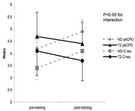
Figure 1. Between-group difference in peak cardiac power output (pkCPO) and cardiac reserve (C-res) pre and post exercise training
Where applicable, experiments conform with Society ethical requirements.