
Acute exposure to inspiratory hypoxia is an established protocol for the induction of neurovascular headache in the otherwise healthy human brain and is responsible for other vegetative symptoms collectively known as acute mountain sickness (AMS). Though brain swelling may play a role, the lack of any consistent relationship between volumetrics and headache intensity suggests that additional intracranial ‘risk’ factors may be involved. The inability to accommodate brain swelling subsequent to insufficient intracranial volume reserve (IVR) may predispose to hypoxic headache (HH) and thus by consequence, AMS (Ross, 1985). To examine this, 20 healthy subjects were examined in normoxia (PRE-NORM), following 16h passive exposure to 12% O2 (HYP) and after 6h recovery in normoxia (POST-NORM). MR-images were acquired on a 1.5T scanner using a standard head coil and T1-weighted gradient-echo sequences were applied across the whole brain to the level of the foramen magnum. Volumetric changes were processed using SIENA software. IVR was calculated as the ratio of brain volume to intracranial volume (BV:ICV). HH was assessed using a visual analogue scale and clinical AMS diagnosed according to established guidelines (Bailey et al. 2005). Ten subjects (50%) were diagnosed with clinical AMS and corresponding headache scores increased more markedly (HYP minus PRE-NORM) compared to the healthier control subgroup (AMS+ve: +42 ± 18 vs. AMS-ve: +15 ± 14mm, P < 0.05, independent samples t test). While the increase in BV was not selectively different (AMS+ve: +8.3 ± 5.1 vs. AMS-ve: +5.7 ± 4.3mL, P > 0.05), IVR was consistently lower in AMS as indicated by an elevated BV:ICV (Fig. 1). These findings implicate IVR as an anatomical risk factor for HH and AMS. The ‘tight-fit’ brain may predispose to mechano-chemical activation of pain-sensitive structures during hypoxic brain swelling.
University College London 2006 (2006) Proc Physiol Soc 3, C37
Oral Communications: The ‘tight-fit’ brain; an anatomical risk factor for hypoxic headache?
Damian Miles Bailey1, Kai Kallenberg2, Stefan Christ2, Alex Mohr2, Robin Roukens3, Elmar Menold3, Thorsten Steiner4, Peter Bartsch3, Michael Knauth2
1. Department of Physiology, University of Glamorgan, South Wales, United Kingdom. 2. Department of Neuroradiology, Georg August University Medical Centre, Goettingen, Germany. 3. Department of Internal Medicine, University of Heidelberg, Heidelberg, Germany. 4. Department of Neurology, University of Heidelberg, Heidelberg, Germany.
View other abstracts by:
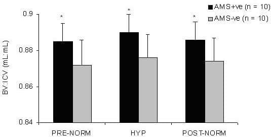
Figure 1. Lower IVR in AMS. Values are mean ± SD; main effect (AMS+ve > AMS-ve P < 0.05 two-way repeated measures analysis of variance); *different vs. AMS-ve (P < 0.05).
Where applicable, experiments conform with Society ethical requirements.