
Recent work on mechanically skinned toad skeletal muscle fibres has shown that depletion of SR Ca2+ by 30 mM caffeine can induce store-operated Ca2+ entry (SOCE, Launikonis et al. 2003). SOCE was detected as a decreased in the fluorescence of fluo-5N, which was trapped within the t-tubules during skinning and subsequent resealing. We have shown previously that (i) in human fibres obtained from malignant hyperthermia (MH) susceptible patients, application of 1 mM halothane induces SR Ca2+ release, which takes the form of a propagated Ca2+ wave and (ii) similar Ca2+ waves can be induced in normal muscle by application of halothane in the presence of reduced cytosolic [Mg2+] (Duke et al. 2004). In this study, we have investigated whether SOCE occurs as a consequence of halothane-induced Ca2+ waves. Rats (200-250 g) were humanely killed. Single extensor digitorium longus (EDL) muscle fibres were mechanically skinned under oil and then perfused with solutions approximating to the intracellular milieu. Exposure of skinned fibres to 50 µM Fluo-5N (pentapotassium salt) resulted in a progressive increase in fluorescence and the emergence of a characteristic sarcomeric pattern (Fig. 1 left), detected using confocal microscopy. Subsequent treatment with saponin abolished the sarcomeric pattern, suggesting that the dye accumulates within the resealed t-system, rather than the SR (not shown). Similar results were obtained in 6 other preparations. In the presence of 0.1 mM [Mg2+], introduction of 0.2 mM halothane resulted in a Ca2+ wave, apparent as localised sarcomere shortening (not shown). The wave was followed by a sustained decrease in fluo-5N fluorescence, consistent with Ca2+ depletion of the resealed t-tubules (Fig. 1 right). Similar results were obtained in 6 other preparations. The t-tubules re-loaded with Ca2+ over 5-10 min and this process could be accelerated by raising the cytosolic [Ca2+] (not shown). In conclusion, the entry of fluo-5N into the resealed t-tubules may occur via anion transporters, which have been implicated in extrusion of fluorescent dyes from the cytosol in intact cells. The decrease in fluo-5N fluorescence following the halothane-induced Ca2+ wave suggests that SR Ca2+ depletion is sufficient to activate SOCE. If SOCE occurs during MH, then Ca2+ entering the cell via this mechanism might contribute to the sustained activation of the myofilaments.
University College London 2006 (2006) Proc Physiol Soc 3, PC107
Poster Communications: Store-operated Ca2+ entry following halothane-induced Ca2+ waves in mechanically skinned rat muscle fibres
Adrian M Duke1, Derek Steele1
1. Institute of Membrane and Systems Biology, University of Leeds, Leeds, North Yorkshire, United Kingdom.
View other abstracts by:
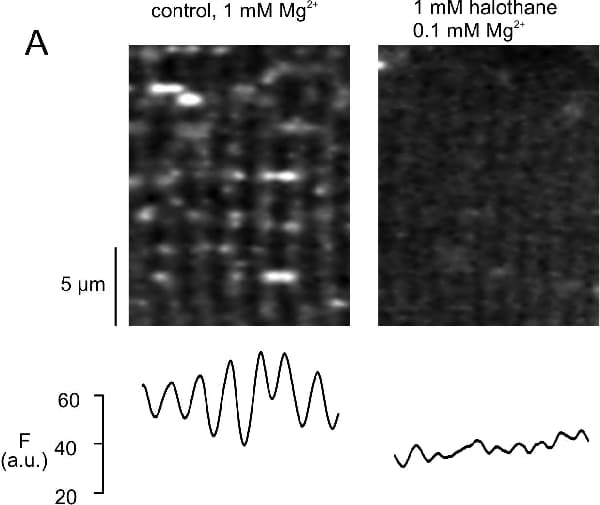
Figure 1 Confocal image of an EDL fibre after 15 min exposure to fluo-5N (left). The same fibre is shown 1 min after addition of 0.1 mM Mg2+ 0.1 mM halothane (right).
Where applicable, experiments conform with Society ethical requirements.