
There is mounting evidence that multi-drug resistance proteins play an important role in fetal protection from drugs and xenobiotics in maternal plasma. The role of these proteins may be of particular importance early in gestation when fetal susceptibility to teratogens is greatest. Here we examine the expression and localization of two multi-drug resistance proteins, P-gp and BCRP, in human placenta during the first trimester as compared to term. First trimester (1 each at 6, 8, 9, 11 and 12 weeks) and term placentas (n=4) were collected and villous fragments dissected, fixed in 4% formaldehyde, paraffin embedded and 4µM sections cut. The sections were de-waxed in xylene and endogenous peroxidase activity blocked using 0.5% hydrogen peroxide in methanol. For BCRP, antigen retrieval was then performed using 10mM sodium citrate buffer. Following this all sections were blocked for 10 min using DAKO protein block and incubated with primary antibody: P-gp Clone F4 (1:20) or BCRP BXP-21 (1:50) overnight at 4°C. A biotinylated secondary rabbit anti-mouse antibody was then applied for 1 hour at room temperature (1:200), followed by 0.5µg/ml avidin peroxidase in 0.125M TBS. The sections were then placed in a bath of 0.05% diaminobenzidine tetrahydrochloride dehydrate (DAB) with 0.015% hydrogen peroxide for 5 min. Methyl green (0.25%) was used as counterstain. Negative controls were processed in exactly the same way, but TBS replaced the primary antibody during the 4°C incubation. Sections were viewed using a Leitz Dialux 22 microscope fitted with a Sony DXC930P camera. All samples for each antibody were processed simultaneously to allow comparison of staining intensity at different gestations. At term both proteins were located predominantly on the microvillous membrane of the syncytiotrophoblast whilst in all first trimester samples a broader distribution throughout the syncytium was observed (Fig. 1). In each first trimester sample staining was markedly heavier when compared with the four term samples, suggesting higher expression of these efflux proteins earlier in gestation. This correlates well with observations of decreasing placental RNA expression for P-gp over gestation (Sun et al. 2005) but conflicts with reports of unchanged RNA expression for BCRP (Mathias et al. 2005). Further quantitative determinations are required to clarify this. A higher functional expression of P-gp and BCRP in the first trimester placenta could act to maintain low levels of xenobiotics in the first trimester fetus thereby giving increased protection from xenobiotic insult during this critical period of development.
University College London 2006 (2006) Proc Physiol Soc 3, PC202
Poster Communications: Gestational changes in expression and localization of P-glycoprotein (P-gp) and breast cancer resistance protein (BCRP) in human placenta
Diane Elizabeth Atkinson1, Susan L Greenwood1, Colin P Sibley1
1. Division of Human Development, The Medical School, University of Manchester, Manchester, United Kingdom.
View other abstracts by:
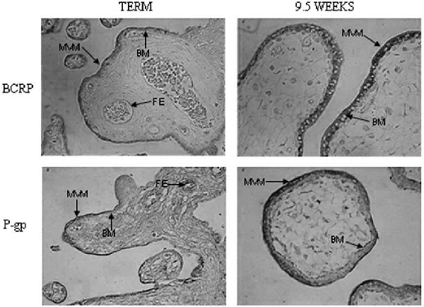
Figure 1. Expression and localization of BCRP (BXP-21) and P-gp (F4) in human placenta at term (representative of 4 samples) and 9 weeks gestation. A similar distribution is seen in all first trimester samples (magx400 MVM-microvillous membrane BM-basal membrane FE-fetal endothelium).
Where applicable, experiments conform with Society ethical requirements.