
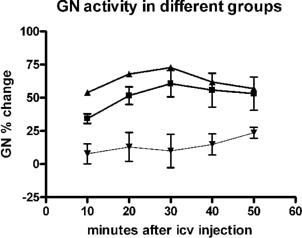
Peripherally released cytokines enhance sympathetic drive to a number of targets (1). One probable mechanism is through the activation of PGE (EP) receptors following cytokine-induced prostaglandin release from endothelial cells of the cerebral circulation. As cytokine levels are raised in systemic inflammatory states that increase cardiovascular risk, such a mechanism may contribute to hypertension, raised sympathetic activity and other physiological disorders (1,2). Here the actions of ICV PGE1 are examined upon population of muscle vasoconstrictor (MVC) sympathetic activity recorded from a gastrocnemius nerve (GN). Urethane-anaesthetised male rats (N=9; initial dose, 1.3 g kg-1, I.P., supplemented with 5-10 mg I.V. as required) were positive pressure ventilated with oxygen-enriched air and maintained in central apnoea (3). Diaphragmatic EMG was recorded and blood gases monitored to assess central respiratory drive. Glass suction electrodes were used to record mvc and cutaneous vasoconstrictor (CVC) activities simultaneously from a GN and the ipsilateral tibial nerve plantar branch (TNp), respectively (filtered, 80-1000 Hz; rectified and smoothed (see 3)). Either PGE1 (500 ng in 5 µl ACSF) or vehicle was administered ICV, as previously described (see 4). Of three groups of animals, 2 groups received PGE1; one group was vagotomized (A: N=2) and the other additionally sinoaortic denervated (B: see 3: N=4). Animals in the time matched control group (C; N=3) received vehicle only and were vagotomized and sinoaortic denervated. MVC data were analysed only where CVC activity increased following PGE1 (positive control; see 4). Ten minutes following the administration of PGE1 the change in MVC activity seemed greater than that seen in the group receiving vehicle (Fig. 1). The effect appeared to be sustained for 50 min following PGE1. Although the sample size in this preliminary study precluded statistical analysis on differences between treatments at each time point, the mean values from all time points (n=5) in each treatment group were compared (1-way ANOVA): MVC percentage change in each of the PGE1-treated groups (A = 51±5%; B = 63±3%) was significantly different from that in the vehicle control (14±3%, p < 0.0001). BP and heart rates in groups A and B were similarly significantly different from C (mean BP 114, 128 and 86 mm Hg respectively; mean heart rate 505, 528 and 460 beats/min, respectively; n=5, p < 0.001) These preliminary studies are consistent with the idea that activation of CNS EP receptors can lead to an increase in MVC activity which if maintained may contribute to cardiovascular disease.
University College London 2006 (2006) Proc Physiol Soc 3, PC68
Poster Communications: Possible involvement of central prostaglandins in elevated muscle vasoconstrictor activity associated with systemic inflammatory states
Michael P Gilbey1, Chunhua Huang1
1. Physiology, UCL, London, United Kingdom.
View other abstracts by:
Where applicable, experiments conform with Society ethical requirements.