
Chronic restraint stress (CRS), in which rats are held for periods of 6 h/day for up to 21 days, induces raised corticosteroid levels and may result in cognitive impairment. It causes a variety of morphological changes in hippocampal areas, including loss of dendritic arborisation but, at least in area CA3, no significant neuronal loss. We have examined the effects of 21 days of CRS on the ultrastructure of synapses, dendrites and spines in rat hippocampal areas CA3 and CA1. In order to ensure that our data were not biased by volume changes the Cavalieri method was used to assess the effects of CRS on hippocampal volume. Following CRS, the mean total hippocampal volume decreased by ~15%, and for dorsal CA1, the volume was reduced by ~16%; in contrast, in CA3 there were no significant volume differences between control and stressed rats. These data were used to correct density measurements in subsequent studies. At the ultrastructural level, using unbiased 2-D stereology, we found that there is a loss of simple unperforated synapses in striatum lucidum of CA3, which can be reversed rapidly by spatial learning (Sandi et al. 2003). To determine the real nature of synaptic and dendritic changes we used full 3-dimensional reconstruction (software from Fiala and Harris: http://synapses.bu.edu) of these structures (Stewart et al. 2005). Up to 150 serial sections (~50-70nm in thickness) were taken for each of the 3-D reconstructions of dendritic segments (~20 μm in length) of CA3, to show whole thorny excrescences (the spine complexes found in CA3), and postsynaptic densities (PSDs) on the thorns, for each of four functional animal states: (i) unrestrained controls, (ii) restrained, (iii) water maze-trained and (iv) water maze-trained, following restrain stress. A segment of dendrite from CA3 with thorny excrescence is shown in Fig. 1. Note the thorns with post-synaptic densities (PSD) in red, and also a single mushroom spine, also with a PSD (scale bar, 5μm). Following 21 days of chronic restraint stress (CRS), our 3-D data show that in CA3 there is a decrease in volume of thorns and a retraction of thorns in CRS compared with unrestrained rats. However, there is no change in the number of thorns per thorny excrescence. CRS induces a decrease in both endosome content and coated vesicles in thorns, processes which are substantially reversed 24 h following water maze training and indicating most probably in relation to the endosome changes, altered receptor recycling. However, in CA1 changes in 3-D structure are not so marked (Donohue et al. 2006). Our data from CA1 (so far only with stressed and control rats), have shown no quantitative changes in spine parameters between groups but significant increases in PSD membrane surface area (>36%) and in PSD volume (>60%) in stratum lacunosum moleculare of the CRS group. A highly significant overall increase in the ‘PSD surface area/spine surface area’ ratio also occurs in the CRS group (>27%). Overall these data indicate that as a result of CRS there is a highly selective structural remodelling of dendritic and synaptic contacts within CA3 and CA1, which for CA3 can be rapidly reversed by a behavioural training task, demonstrating the remarkable neuroanatomical plasticity of CA3.
University of Bristol (2007) Proc Physiol Soc 5, SA13
Research Symposium: Dendritic and synaptic remodelling in mammalian hippocampus following stress
Michael G. Stewart1
1. Biological Sciences, The Open University, Milton Keynes, United Kingdom.
View other abstracts by:
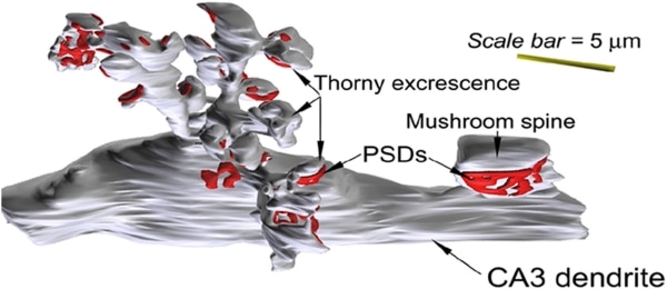
Figure 1. A 3-dimensional reconstruction from 150 serial ultrathin sections of a 20 μm segment of dendrite from CA3 with thorn excrescences. Note that the thorns show post-synaptic densities (PSD) in red and there is also a single mushroom spine with a PSD (scale bar 5μm).
Where applicable, experiments conform with Society ethical requirements.