
The effects of acidosis on the electrophysiology of the sinoatrial (SAN) and atrioventricular (AVN) nodes were investigated. Hearts from adult male New Zealand White rabbits were excised under terminal anaesthesia. In one series of experiments (n=6), optical mapping was used to image left ventricular activation in Langendorff-perfused rabbit hearts stained with the voltage-sensitive dye RH237 and paced from the right atrium (RA). Left ventricular epicardial activation was recorded at 16×16 sites in a 2x2cm square. In another series (n=11), SAN cycle length (CL) and AVN conduction were studied in an isolated right atrial/AVN preparation, containing the triangle of Koch and crista terminalis, superfused with Tyrode’s solution at 37°C. Extracellular electrodes recorded atrial and His bundle signals during sinus rhythm or pacing from the crista terminalis. Atrio-Hisian (AH) interval, Wenckebach cycle length (WCL) and AVN functional (FRP) and effective (ERP) refractory period were derived from pacing protocols. Data were obtained at pH 7.4, 6.8 and 6.3 (Table 1). Comparisons of values at each pH increment were made using a paired t-test, and a two-tailed P-value of < 0.05 was considered statistically significant. In intact hearts, the time to earliest ventricular activation (TEact) following RA pacing prolonged as PCL shortened, consistent with delay originating at the AVN. Reduction of perfusate pH resulted in significant and reversible prolongation of TEact. In the isolated preparation, the spontaneous SAN CL lengthened with reduction of pH. There was reversible prolongation of the AH interval and WCL during acidosis, with significant prolongation of AVN FRP and ERP. In conclusion, acidosis prolongs the time to earliest ventricular activation from RA pacing due to a significant increase in AVN delay. Furthermore, acidosis prolongs the refractory period of the AVN and slows the SAN firing rate.
University of Manchester (2007) Proc Physiol Soc 8, PC12
Poster Communications: Acidosis delays conduction through the atrioventricular node
A. M. Nisbet1, M. Craig1, N. L. Walker1, F. L. Burton1, S. M. Cobbe1, J. Hancox2, C. Orchard2, G. L. Smith1
1. BHF Glasgow Cardiovascular Research Centre, University of Glasgow, Glasgow, United Kingdom. 2. University of Bristol, Bristol, United Kingdom.
View other abstracts by:
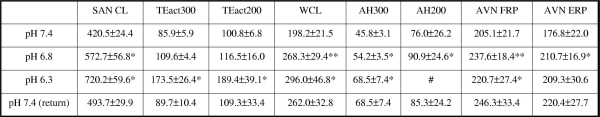
Table 1 Data expressed as mean ± SEM (ms). TEact300 - TEact at PCL 300ms; TEact200 - TEact at PCL 200ms; AH300 - AH at PCL 300ms; AH200 - AH at PCL 200ms. *p<0.05, **p<0.01, # At pH 6.3 data was not included in analysis due to complete heart block at PCL 200ms in 9 of 11 samples.
Where applicable, experiments conform with Society ethical requirements.