
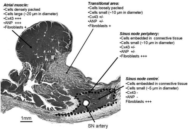
We have shown that functionally and morphologically the rabbit sinus node (SN) is an extensive structure – it extends from the superior (SCV) to inferior (ICV) caval vein (1,2). It has also been shown that in the human, the leading pacemaker site can occur anywhere between the SCV and ICV (3). Therefore, the aims of this study were to determine if morphologically the human SN is a more extensive structure than previously thought and to investigate cell types within and around the SN. Four healthy human SN/atrial specimens (not used for transplantation from Queensland Heart Valve Bank) were used. Serial frozen tissue sections were stained for histology (with Masson’s trichrome stain) to locate the SN. Adjacent sections were immunolabelled with various antibodies to distinguish different population of cells within and around the SN. Cx43 (major connexin in the working myocardium) and ANP (atrial natriuretic peptide) were used as negative markers of nodal tissue; caveolin3 and vimentin were used as markers of cardiac cells and fibroblasts respectively. Western blotting was used to test the specificity of the antibodies. Scion Image was used to measure the intensity of immunofluorescence signal captured by confocal microscopy. With these techniques four different regions in each tissue section were identified: atrial muscle (AM), transitional area (TA), periphery (SNp) and centre (SNc) of the SN. Summary of data is shown in Fig. 1. The AM is characterised by large densely packed myocytes with little connective tissue and fibroblasts, but high expression of Cx43 and ANP. The TA is characterised by small loosely packed myocytes that contains a mixture of Cx43 and ANP positive and negative cells. The SNp, like TA, also contains a mixture of Cx43 and ANP positive and negative cells, its cells are smaller than AM cells, but similar to TA cells. However, unlike TA cells, SNp cells are embedded in network of connective tissue. The SNc is characterised by much smaller cells embedded in network of connective tissue, contains large number of fibroblasts and little or no Cx43 and ANP. The location and size of the SN, in our study, is in agreement with previous studies (e.g. 4). However, the TA extended further into the AM of the terminal crest and extended beyond the SN towards the ICV. We conclude that the SN is more extensive structure than previously thought, and the large transitional area (which contains a mixture of atrial and nodal cells) could explain the large variation in the position of the leading pacemaker site.
University of Manchester (2007) Proc Physiol Soc 8, PC18
Poster Communications: Histological and immunohistochemical characterisation of cells types in different regions of the human sinus node
N. J. Chandler1, P. Molenaar2, S. Birchall1, H. Musa1, V. Sharma3, D. C. Sigg3, M. R. Boyett1, H. Dobrzynski1
1. Division of Cardiovascular and Endocrine Sciences, University of Manchester, Manchester, United Kingdom. 2. University of Queensland, Queensland, QLD, Australia. 3. Medtronic Inc., Minneapolis, MN, USA.
View other abstracts by:
Where applicable, experiments conform with Society ethical requirements.