
The sympathetic nervous system accelerates heart rate via β adrenergic receptors (βARs) on sinoatrial node (SAN) myocytes. Knockout studies suggest that the β1AR subtype mediates adrenergic control of heartrate in the mouse.1,2 Hyperpolarization-activated cyclic nucleotide sensitive (HCN) channels are thought to be among the downstream effectors for this chronotropic effect. HCN4 is the main HCN channel isoform in the SAN.3 We hypothesized that close association between β1ARs and HCN4 channels would facilitate the rapid and efficient signaling that is critical for sympathetic control of heartrate. Here we have used immunofluorescent confocal microscopy to examine the distribution of β1ARs and HCN4 channels on SAN cells. Adult male mice were anesthetized with isoflurane and euthanized according to a University-approved protocol. SAN tissue was digested in a Ca2+-free enzymatic cocktail and cells dissociated with a fire polished glass pipette. Following gradual reintroduction of Ca2+, cells were plated onto poly-L-lysine coverslips, fixed with 4% paraformaldehyde, and permeabilized with 0.2% Triton X-100. Rabbit polyclonal primary antibodies against HCN4 (Alomone) or β1AR (Santa Cruz) were applied at 4oC overnight, and visualized with an Alexa488-conjugated goat anti-rabbit secondary antibody (Molecular Probes). Optical sections (0.5 μm) were acquired with a 100x oil objective on a Leica TCS SP2 laser scanning confocal microscope. Antibody specificity was confirmed by Western blotting of lysates from mouse atria or from CHO cells transfected with β1AR, β2AR, HCN2, or HCN4. Other controls included antibody preadsorption with antigenic peptides and omission of primary antibodies. All data represent at least three independent experiments. Both HCN4 and β1AR were conspicuously located on SAN cell membranes at discrete sites that appeared to be regions of contact between adjacent myocytes. HCN4 and β1AR immunofluorescence was seen in lateral membrane regions and at the ends of the cells. These patterns was present in both larger, presumed “transitional,” cells and in smaller, more slender “primary” cells. Additional HCN4 fluorescence was seen along the entire cell membrane, and additional β1AR fluorescence was seen in puncta across the cell surface. In pairs of joined cells, there was marked β1AR or HCN4 fluorescence at the cell-cell interface (Figure 1). These data suggest that HCN4 and β1ARs are both localized at intercellular junctions between myocytes in the SAN.
University of Manchester (2007) Proc Physiol Soc 8, PC3
Poster Communications: Subcellular localization of β1 adrenergic receptors and HCN4 channels in murine sinoatrial myocytes
P. Akella1, J. Vega1, C. Proenza1
1. Physiology & Neurobiology, University of Connecticut, Storrs, CT, USA.
View other abstracts by:
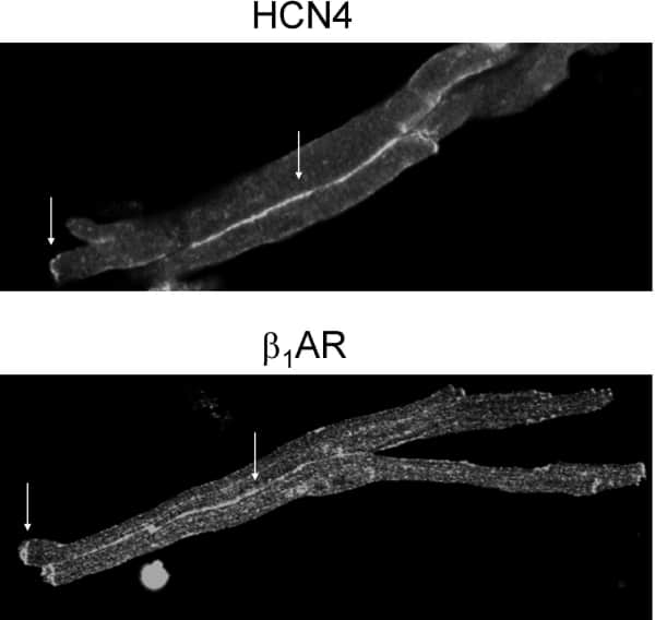
Figure 1. Pairs of sinoatrial myocytes labeled with anti-HCN4 (top) or anti-β1AR (bottom) primary antibodies and an Alexa488-conjugated secondary antibody. Arrows indicate immunofluorescence at the ends of myocytes and at the interface between the cells.
Where applicable, experiments conform with Society ethical requirements.