
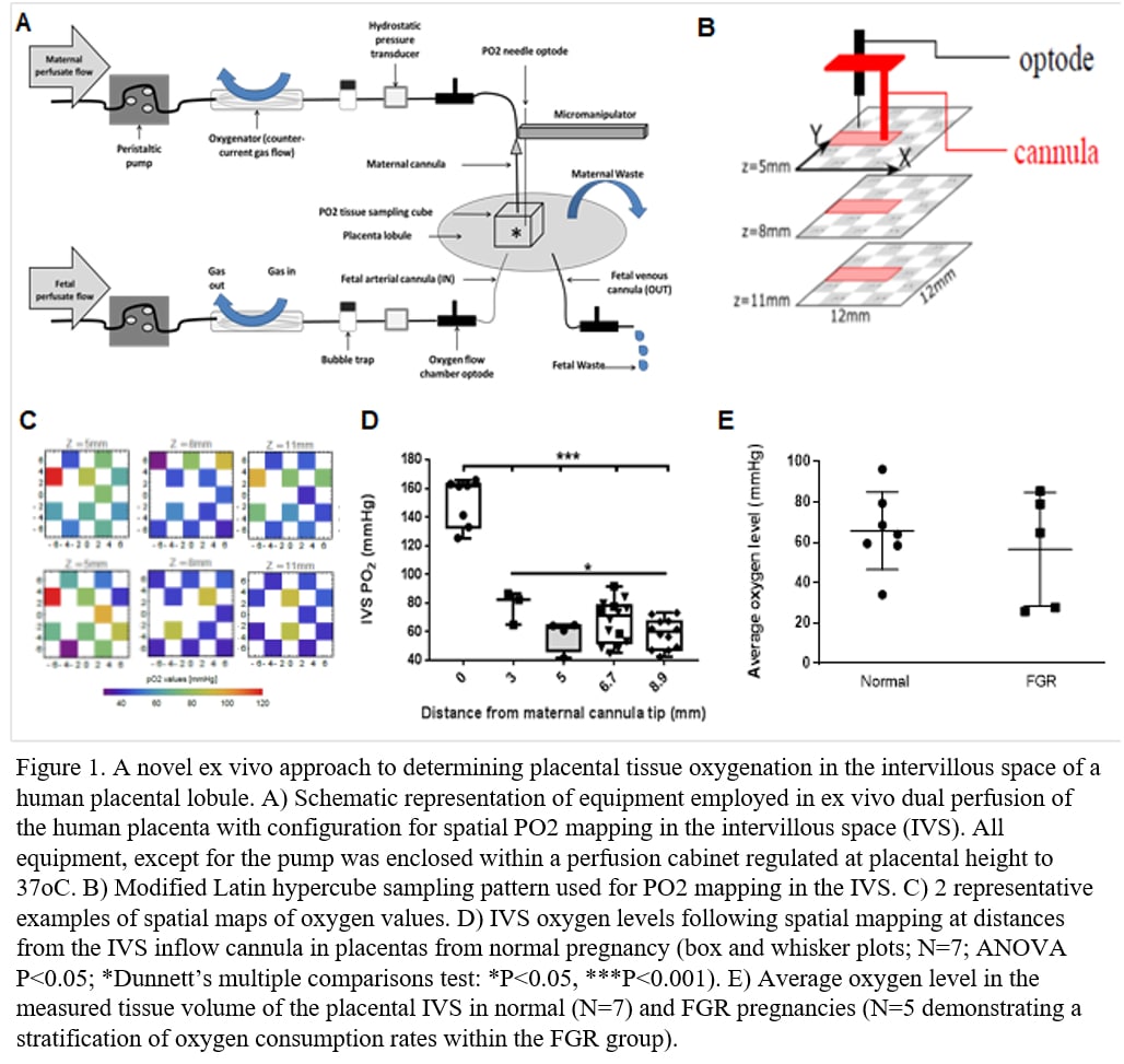
Introduction: Fetal growth restriction (FGR) is a global problem affecting 3-7% of pregnancies worldwide(1). Fetal growth depends on adequate oxygen supply(2). Understanding oxygenation at significant depths within organs is an unmet challenge, constrained by tissue accessibility and spatial resolution, preventing an understanding of metabolic dysregulation in the healthy and diseased state. This is especially challenging in the placenta with its complex anatomy and dual blood flows(3). Methods: Here we overcome this problem using novel methodology based on ex vivo perfusion of the human placenta employing high-resolution 3D oxygen-mapping of the intervillous space(IVS). Tissues were acquired following informed written consent from women attending St Mary’s Hospital, Manchester, UK. Normal singleton pregnancies above the 20th centile for fetal weight(N=7) and diagnosed cases of FGR in pregnancy and a live birth weight below the 5th centile (FGR group, N=5) were recruited. Ex vivo perfusion was performed as previously described(4) adapted through using only one maternal side cannula, replicating a maternal single spiral artery and perfusing the IVS (Figure 1A). An oxygen-sensing optode and oxygen monitor measured oxygen readings in a Latin hypercube arrangement, predicted to optimise data collection for in silico modelling, capturing a 1.2×1.2×0.6 cm3 volume of the placental lobule (Figure 1B). Results: There was a significant and consistent loss of oxygen with increased distance from the cannula in the normal group, equating to 90±34 mmHg at 8.9mm away from the cannula tip, or a loss of 10.1± 3.8 mmHg/mm (ANOVA P<0.05; *Dunnett’s multiple comparisons test: P<0.05; Figure D). Further interrogation showed 47±13% of the oxygen provided was lost in the first 3mm from the source. Average IVS PO2s revealed high variance within both groups (63.99, 58.48-79.41 mmHg and 64.76, 26.83-82.17 mmHg, median and interquartile rages, normal and FGR, respectively; Figure 1E) but in the FGR group there was a clear stratification at the extremes of normality although this has not yet reached significance (P=0.38, F test) (Figure 1E). Discussion: The stratification of oxygen gradients within the FGR group may be linked to the severity of the disease. The results gained form these methods have the potential to transform our understanding of spatial oxygen metabolism in the human placenta, providing a ground-breaking insight into the way oxygen diffusion, blood flow and microanatomy combine, stimulating further in vivo diagnostic development to assist in the management of pregnancy problems. The technology we report can be tailored to gain insight into spatial aerobic metabolism in a range of perfused tissue types.
Physiology 2019 (Aberdeen, UK) (2019) Proc Physiol Soc 43, C075
Oral Communications: A novel ex vivo and in silico approach to determine placental tissue oxygenation in normal and fetal growth restricted pregnancies
G. A. Nye1,2, A. Erlich3, E. Johnstone2, R. Lewis4, I. chernyavsky3,2, P. Brownbill2
1. Chester Medical School, University of Chester, Chester, Chester, United Kingdom. 2. Maternal and fetal health, university of manchester, Manchester, Manchester, United Kingdom. 3. School of Mathematics, University of Manchester, Manchester, United Kingdom, United Kingdom. 4. Faculty of Medicine, University of Southampton, Southampton, United Kingdom.
View other abstracts by:
Where applicable, experiments conform with Society ethical requirements.