
Introduction: Cardiac reflexes originating from sensory receptors in the heart ensure blood supply to all tissue and organs in the face of constantly changing demands. Atrial receptors are mechanically sensitive vagal afferents which relay to the medulla and hypothalamus, affecting vasopressin release and renal sympathetic activity. Although the mechanism of volumetric mechanotransduction has not been studied before, vesicle recycling is an important component in other mechanotransduction systems (1). Two distinct sensory endings for atrial receptors have been localised to the subendocardial space using methylene blue staining or anterograde tracing (2) and these are referred to as end-nets and flower spray endings. To map the distribution of atrial receptors in the subendocardial space, we have double-labelled rat right atrial whole mounts [neurofilament (NF) & synaptic-vesicle protein 2 (SV2)] and generated high resolution maps of the rat subendocardial neural plexus. Methods: In accordance with EU directive 2010/63 and local ethical committee approval, 5 female wistar rats (150-350g) were euthanised by cervical dislocation following isoflurane anaesthesia (5%). The heart was rapidly removed and the right atrium was isolated, pinned flat on silicone with the endocardial surface exposed and fixed & permeabilised in precooled methanol (-20 °C, 30 mins). Subsequently the tissue was blocked and incubated overnight in a humidity chamber at 4°C with primary antibody [1:500 chicken anti-neurofilament (Abcam, ab72996), 1:25 mouse anti-synaptic-vesicle protein 2 (Hybridoma Bank)]. Primary antibodies were rinsed off with blocking solution prior to secondary incubation in a light-protected humidity chamber overnight at 4°C [1:250 goat anti-chicken IgY Alexa Fleuor®568 (Abcam, ab175477), 1:100 goat anti-mouse IgG-FITC (Sigma F8771)]. Samples were imaged using an apochromatic objective lens. Images were stitched using the Pairwise stitching function included in Fiji (ImageJ) (3). Results: The subendocardial space of the right atrium was richly innervated in all specimens and a recurrent hourglass-like arrangement of axons was present in the posterior atrial wall. Extensive end-net formations were visualised at the cavo-atrial junctions and interatrial septum. NF and SV2 antibodies differentially stained axons with NF labelling larger axons while SV2 labelled smaller axons. Nets of SV2 labelled endings were coincident with NF labelled nets particularly at the cavo-atrial and mid-atrial regions Conclusion: A smaller set of vesicle filled terminals extends from the larger and previously described end-nets. The study has provided an anatomical basis by which atrial deformation (atrial filling and contraction) can be communicated to the brain.
Physiology 2019 (Aberdeen, UK) (2019) Proc Physiol Soc 43, C102
Oral Communications: Mapping atrial receptor distribution in the right atrial subendocardial neural plexus of the rat
T. Campbell1, F. Shenton2, S. Pyner2, J. F. Jones1
1. School of Medicine, University College Dublin, Dublin, Ireland. 2. Department of Biosciences, Durham University, Durham, United Kingdom.
View other abstracts by:
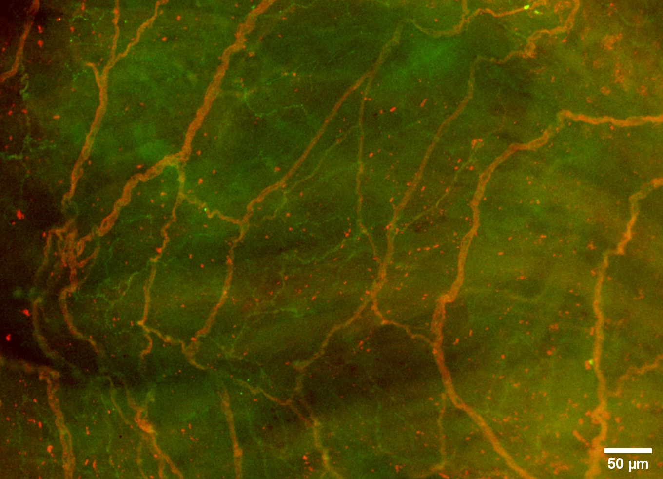
Figure 1: Subendocardial end-net of the inferior venous-atrial junction. Epifluorescent imaging of rat right atrial whole mount showing large red end-nets (neurofilament heavy chain) and smaller green nets (synaptic vesicle protein 2). The presence of synaptic vesicle protein 2 indicates the presence of vesicles. No varicosities are observed in the synaptic vesicle protein 2 net which suggests that these are sensory in nature. No flower spray endings are present. Images were captured using an apochromatic plan objective lens. Images were contrast enhanced and overlayed using Fiji (ImageJ).
Where applicable, experiments conform with Society ethical requirements.