
Induction of an oscillatory flow pattern or increasing retrograde flow has been associated with proatherogenic effects in endothelial cell culture models1. Retrograde flow in peripheral arteries is also known to increase with ageing2. The flow pattern at any point depends on pressure difference across that point3. One of the factors which can influence the pressure waveform, is the velocity of propagation of the reflected waves, which are directly affected by vascular stiffness. The aim of the study was to investigate the association between vascular stiffness and brachial artery flow profiles in patients of myocardial infarction. Patients of recent (<6months) myocardial infarction (n= 25) were recruited from the Department of Cardiology, AIIMS, New Delhi. Flow profile was assessed at the brachial artery using Pulsed wave doppler (M7, MindRay) with simultaneous acquisition of the diameter in duplex mode for estimation of shear rate (Shear Rate = 4*Velocity/Diameter). Oscillatory shear index was calculated using the formula: Retrograde shear rate/ (Anterograde shear rate + Retrograde shear rate). Regional vascular stiffness was assessed by measuring the Pulse Wave Velocity (PWV) of the carotid-radial (cr) arterial segment using applanation tonometry (SphygmocorÒ). Carotid-Intima-Media Thickness (CIMT) was measured using B-mode ultrasound (M7, MindRay) for estimation of subclinical atherosclerotic burden. Carotid artery wall properties were derived from carotid artery diameter changes during the cardiac cycle for calculation of compliance and Incremental Elastic modulus (Medical Imaging applications Carotid Analyser). A significant positive correlation was observed between cr-PWV and oscillatory shear index (OSI) (r = 0.44, p = 0.01) and between far-wall CIMT and OSI (r = 0.45, p = 0.01). A significant negative correlation was seen between Carotid compliance and OSI (r = -0.35, p = 0.03) and a significant positive correlation was observed between Incremental Elastic modulus and OSI (r = 0.38, p = 0.02). Both OSI and retrograde flow correlated positively with Age (r = 0.49, p = 0.004 and r = 0.36, p = 0.03). The current study highlights the factors associated with the atherogenic-oscillatory flow pattern. Preliminary results suggest a potential role of increase in vascular stiffness of the corresponding regional arterial segment and a decrease in carotid artery compliance. Additionally, the study shows an association between oscillatory shear pattern and macroscopic sub clinical atherosclerosis. A decrease in carotid artery compliance would alter the characteristic plateau pattern of the central pressure waveform to a sharper wave with a steep decline in pressure. This would be further augmented by increase in regional arterial stiffness leading to an amplification of the reflected waves. The net effect of the altered pressure waves would result in development of an oscillatory shear pattern in patients of myocardial infarction.
Physiology 2019 (Aberdeen, UK) (2019) Proc Physiol Soc 43, C109
Oral Communications: Association between vascular stiffness and oscillatory flow pattern in patients with recent Myocardial Infarction
S. Badhwar1, D. S. Chandran1, A. K. Jaryal1, R. Narang2, C. Patel3, K. K. Deepak1
1. Physiology, All India Institute of Medical Sciences, New Delhi, New Delhi, Delhi, India. 2. Cardiology, All India Institute of Medical Sciences, New Delhi, New Delhi, Delhi, India. 3. Nuclear Medicine, All India Institute of Medical Sciences, New delhi, New Delhi, Delhi, India.
View other abstracts by:
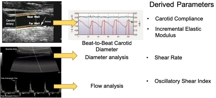
Figure 1. Representative Images of carotid and brachial artery for analysis
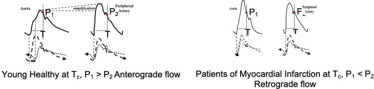
Figure 2. Proposed mechanism for alteration in waveforms leading to oscillatory flow in Myocardial Infarction
Where applicable, experiments conform with Society ethical requirements.