
Physiology News Magazine
Visualising cell biology: We’ve an app for that!
How I am incorporating molecular visualisation into my teaching
Features
Visualising cell biology: We’ve an app for that!
How I am incorporating molecular visualisation into my teaching
Features
John Barrow, University of Aberdeen, UK
https://doi.org/10.36866/pn.116.28
Physiology depends on interactions between molecules. With the advent of modern visualisation methods, John Barrow, recipient of the David Jordan Teaching Award, discusses some of the ways that these often abstract molecular processes can be viewed to enhance the student learning experience. The David Jordan Teaching Award enables you to carry out a piece of educational research or to develop an educational resource that is relevant to physiology.
Visualisation for learning
Physiology depends on interactions between molecules. Histones wrap around DNA to package genes into the nucleus, and myosin and actin filaments slide past each other to make muscles contract. Biology is inherently dynamic, and yet most teaching materials (e.g. textbooks, lecture slides, etc.) show static representations of physiological processes. For those students who can think visually, this kind of static imagery can work as they are able to imagine what these processes would look like. However, for many it equates to a set of abstract ideas (e.g. “molecule A is converted into molecule B”, or “molecule X interacts with receptor Y”) that make the biology challenging to visualise and understand. With the advent of modern visualisation methods there is an opportunity to develop immersive and engaging material for students that will address some of the challenges they face when it comes to their learning. Indeed, if a student can engage with visual representations, then they would have the ability to think visually about dynamic processes (Herráez and Costa, 2013).
Changing with the times
My teaching is largely based on metabolism and biochemical pathways. When I studied biochemistry as an undergraduate my lecturers took a rote learning approach. Fast forward a number of years, and when I had the opportunity to teach the same subject I jumped at the chance, but I was keen to do it differently. My main goal was to make it more accessible for all of our life sciences students, as many of them encounter biochemistry and molecular biology in their foundation years but will not go on to study the subjects at an advanced level.
When I started teaching biochemistry it was my first teaching role, so I adopted lectures from a retiring academic and initially didn’t change them very much. After a couple of years of teaching the material in much the same way as I had been taught, I began looking into ways of making the material more engaging and stimulating for students, as well as finding ways of making the concepts as digestible as possible. I began the process of switching from a rote teaching style by incorporating more visual components into my classes. This initially involved adding in videos of metabolic processes so that students could see them in action. From class feedback and talking directly with students, it was clear that these helped some students to understand the processes that had been presented statically in my lectures and their textbooks. But for me this was not enough as the imagery being shown was still a two-dimensional approach as it was being projected onto the lecture theatre wall. I wanted something that allowed students to fully interact with how the processes were actually working in their cells and bodies.
My initial foray into making classes more interactive was the use of 3D-printed protein models. My intention was to show the regions on the proteins that were important to their function, but this wasn’t viewed favourably by students. They really liked the idea of seeing the proteins, but because the protein models are made from hard plastic it is difficult to convey the dynamic nature of the processes being taught. This is where digital visualisation comes in, as it offers a highly visual approach, but also provides interactivity to get students fully engaged in their studies.
Augmenting reality
There are several modern approaches to visualisation including virtual reality (VR) and augmented reality (AR). VR can be defined as a fully visualised world that a user can interact with via a headset. AR can be defined as digital visualisations being overlaid on the physical world around the user that usually require a mobile device such as a tablet or smartphone (for a really good guide see Garcia-Bonete et al., 2019).
Both VR and AR have their pros and cons. VR allows for a fully immersive user experience via a headset, but it is usually a single user experience as others around that individual are not viewing the experience in the same way. AR allows users to see the processes on a device but not in a fully immersive way, which then allows for more interactivity between users. Because of this, I decided to use AR as it allows students to interact and see the visualisations, but also provides them the opportunity to work together in class.
How do you make visualisations?
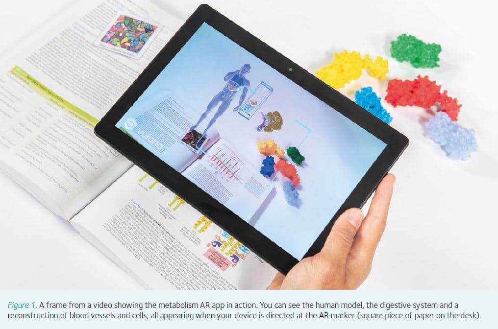
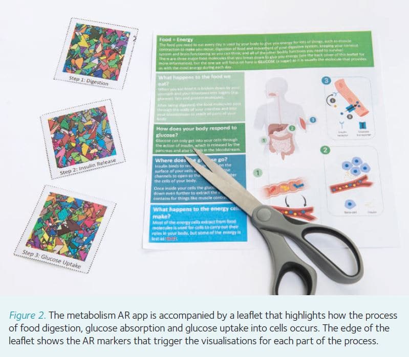
Prior to receiving the David Jordan Teaching Award, I had created an AR app that shows insulin signalling and glucose absorption for high school pupils. To create this three-stage process showing food digestion, insulin release, and insulin action on muscle, the production of nine different models was required, with most of them being created from scratch and others being purchased from various asset repositories. These models were a human figure (1), a chocolate bar (2), hollow tubes to represent the gut (3) and blood vessels (4), pancreatic (5) and muscle cells (6), food particles (7), and finally glucose (8) and insulin (9) molecules. Once the models have been created, they then need to go through a process of refinement to make them as simplistic as possible without removing too much detail. 3D models are made from a wireframe of polygons, so a curved surface on a model is actually made up of many smaller flat-surfaced polygons joined together in a mesh. The more complex the mesh (i.e. the more polygons), the more challenging it is for a device to display, so to make a model that will be visible and animate smoothly on as many devices as possible requires the polygon number to be as low as possible. This creates a challenge, as reducing polygons makes a model less refined (akin to high-resolution graphics becoming more pixelated), so a lot of time and effort are put into minimising the polygon count whilst keeping the model true to form. Once all of the models have been created and refined, they are then given a surface. Surfaces are created to give each model specific surface colours or patterns, as well as allowing the models to be shaded to give them a more realistic appearance. Surfaced and shaded models are then imported into a game engine to allow the creation of animations and visual effects. Game engines are often used as they allow 3D models to be manipulated and animated with relative ease. For this particular application, the game engine allowed the models to be given textures that gave detail rather than the wireframe providing the details – this process makes the models more compatible with mobile devices that have limited processing and graphical capabilities. Following texturisation of the models, they are then animated using the mechanics of the game engine to code the movement for each model where required.
Challenges of working with digital creators
Making augmented reality content has given me some interesting challenges that have been an unexpected surprise. Most of these challenges centred on trying to explain and describe the processes and how they should be visualised. Working with game design experts can be tricky at times as they are incredibly capable when it comes to creating content, but their understanding of the underlying biology is often basic, as the last time they studied biology was in secondary school. Having individuals who are skilled in visualisation methods but also have a solid grounding in the subject matter would streamline the production of high-quality visualisations. Perhaps the addition of coding and computer-aided design, or creative arts in science curricula is something that could work for some individuals wishing to pursue careers in this area, but that is a different story for another day. In practice, the collaboration of game designers, digital artists and me as the scientist worked because we had a process of careful iterations and beta-testing at each stage of the design process. Once each model, animation or design phase was complete, they were tested and approved, then we moved onto the next one in a highly detailed and structured way.
Applications of visualisation
As described here, there are many applications for visualisation in education settings, but the potential to use this type of technology in other areas is huge. Being able to visualise patient data, from brain scans to doppler ultrasound, could allow patients to see the issues they are facing and allow clinicians to easily explain the problems they are hoping to treat. Other areas where visualisation can be of benefit is in allowing patients to experience procedures they may face – a good example of this is the use of virtual reality apps for children who are undergoing stressful or invasive therapies. These technologies are also being used to help individuals deal with situations, such as those with autism, so that they can begin to train themselves to deal with situations in a safe environment that can simulate real world environments. There are also benefits for scientific research, such as the recent virtual reality tumour visualisations that allow scientists to immersively visualise tumours to gain a greater understanding of the structure and cell types found within the tumour (Walsh, 2018).
For me, being able to use this technology to convey the fundamental principles of processes such as receptor function, enzyme activity, protein–DNA interactions and protein–protein interactions will hopefully prove a positive experience for my students. The app I am currently developing with the David Jordan Teaching Award will highlight the dynamic nature of glycolysis and how this pathway is the gateway to the majority of metabolism. I would encourage any member of the society with an interest in teaching to apply for an award as it really can be transformative to your teaching and the experience of your students, so watch this space for updates!
Acknowledgements
I would like to thank The Physiological Society for their generous support through the David Jordan Teaching Award, which has allowed me to take my ideas for visualisation much further. The work highlighted in this article has been a collaborative project with Imagin3D (imagin3d.co.uk).
References
Garcia-Bonete MJ et al. (2019). A practical guide to developing virtual and augmented reality exercises for teaching structural biology. Biochem Mol Biol Educ 47, 16–24.
Herráez A, Costa MJ (2013). Biochemical visual literacy with constructive alignment: Outcomes, assessment, and activities. Biochem Mol Biol Educ 41, 67–69.
Walsh F (2018). ‘Virtual tumour’ new way to see cancer. [Online] BBC News Health. Available at: bbc.co.uk/news/health-46527235 [Accessed 1 August 2019]