
Physiology News Magazine
Memories of Andrew Huxley at UCL and his contribution to the science of photonic crystals
Membership
Memories of Andrew Huxley at UCL and his contribution to the science of photonic crystals
Membership
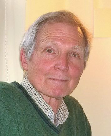
Jonathan A Coles
Visiting Researcher, Institute of Infection, Immunity & Inflammation, University of Glasgow, UK
https://doi.org/10.36866/pn.106.29
The author was a postgraduate at UCL in the late 1960s, and here recalls an aspect of Andrew Huxley’s work which is relatively unknown compared to his famous experiments on the nerve impulse and muscle contraction , to honour the centenary of Huxley’s birth.
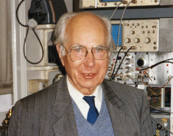
Andrew Huxley at UCL in the 1960s
Huxley had taken the chair of physiology at UCL in 1960, and applied himself conscientiously to his duties. He was very present, punctiliously attending morning coffee and afternoon tea in the common room. At the Christmas party, he would dance the valeta with the technicians and win the drinking-a-yard-of-ale contest. His intellectual presence is illustrated by the advice given to students preparing their first talks: ‘Andrew will probably ask a question, and if he does, there is only one possible answer: Yes, you’re quite right, I should have thought of that.’ The three most closely related departmental heads included Huxley himself, JZ Young in Anatomy, and Bernard Katz in Biophysics, all three outstanding scientists who did experimental work with their own hands, and the atmosphere trickled down. Doug Wilkie and Barbara Banks could be seen crossing by chance in the corridor and haranguing each other with great passion about the thermodynamics of ATP.
It is hard to say how much was due to Huxley’s direct influence, but in addition to subjects like cybernetics and thermodynamics being applied to physiology, many technical aspects of modern electrophysiology were gestating in UCL during his time. He himself motorised the focus on his dissecting microscope, controlling it with his knee. To aim a microelectrode at a selected cell, you need a microscope that can move independently of the micromanipulator, and one early solution was to slide the microscope on a glass plate above a stationary preparation. An early computer, which could just be lifted by a strong person, was acquired for averaging cortical evoked potentials, and people built simple transistor circuits. However, micropipettes were still filled laboriously overnight, as Murakami’s invention in Tokyo of capillaries containing a fused filament had not reached the West. In 1967, Chris Smith and I asked some lecturers what had been the department’s greatest achievement of the year, and the answer was: ‘Kept Huxley’.
The appreciation was not total. People complained about interminable staff meetings, and his lectures were not widely popular. He would demonstrate the compound action potential in frog sciatic nerve in front of about two 200 medical students, and do the calculations, first mentally, then checking on the slide rule he always carried. The medical students asked for him to be taken off, the gist of their argument being that intellectually demanding lectures were an invasion of their safe space, and hearing about the nerve impulse from the person who first quantified it did not make up for this. It was not surprising that Huxley was overjoyed when, in 1969, he was appointed to a Royal Society Research Professorship, and could devote himself to his research.
Biological reflectors
Although Huxley’s main research at UCL was on the mechanism of muscle contraction, he published on a range of subjects. In addition to ‘Proposed mechanism of force generation in striated muscle’ (with Bob Simmons [Huxley & Simmons, 1971]; 1,722 citations), he contributed to a lively exchange in Nature on ‘Sexual activity and beard growth’ (Huxley, 1970: 1 citation), and, prompted by Jack Diamond, did maths on electrotonic potentials. He was also interested in the work of Eric Denton (in Plymouth) and Mike Land (at UCL) on biological reflectors, which work by interference of light in a stack of layers of alternating high and low refractive index. One example is the reflecting scales that camouflage the sides of silvery fish. (If you hold a mirror vertically in the sea, it is hard to see from below because of the way it reflects the sky.) To pass the time on the train home to Grantchester on Wednesday and Friday evenings, Huxley did a mathematical analysis of such one-dimensional multilayer reflectors (Huxley, 1968). The equations for a number of biologically relevant cases were then helpfully plotted by Mike Land (Land, 1972).
Materials that modulate the passage of light by having structure at optical wavelengths are now called ‘photonic crystals’, and multilayer reflectors are a relatively simple, but by no means trivial, case. The first use of ‘photonic crystal’ in an article title seems to have been in 1991 but a search for the term now brings up 46,589 hits, and this does not include spin-offs such as invisibility cloaks. Opticists have been very interested in the diverse kinds of biological reflectors, particularly those in insects. I suspect that one reason for this choice is that a beetle cuticle on a bench top will iridesce, beautifully and odourlessly, for decades, while a herring, say, will not.
Eyeshine in cats
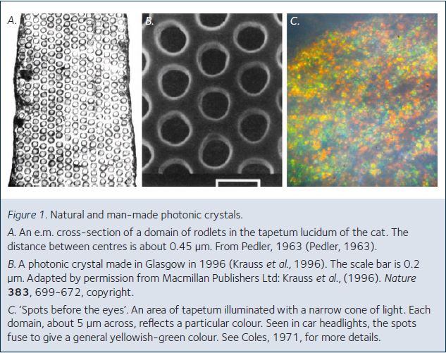
Virtually all nocturnal mammals have a reflecting layer, the tapetum lucidum, behind the retina so that light that escapes absorption on the first passage is not wasted. In Carnivora, the structure is not alternating strata, as in fish scales, but layers of cells each containing arrays of rodlets, as shown for the cat in a 1963 e.m. study by Chris Pedler at the Judd Street Eye Institute (Fig. 1A). For my little MSc project I was told to ‘do something’ with the cat tapetum and to begin by showing Pedler’s images to Huxley. Huxley’s immediate response was that it would give Bragg-type lattice diffraction at optical wavelengths (much as far shorter X-rays are diffracted by atoms in mineral crystals) and that this was interesting and worthwhile demonstrating.
I was provided with a rather nice little brass microscope, and I borrowed Prior manipulators from the teaching lab and reassembled them with various chiralities. It was always a rush restoring them to normal the evening before a practical class. Huxley was tremendously helpful, generous and supportive, but never interfering. He would advise me, for example, to ignore the numbers written on the lamp bulb and to run it at the highest voltage that did not blow it up. He let me use the lathe he kept in his lab, and a Cooke microscope objective that he had had specially modified for water immersion. Of course, we needed cats’ eyes. To get these, I would hang around while people finished cat experiments, which
meant working late at night. Bruce Lynn was one of the worst: he rarely finished an experiment before about 3 am, and only then could I start myself. I learnt that Huxley was often still in his lab at 1 am, and that there was a Czech postdoc who sang magnificently when he thought that everyone else had left.
It was quickly apparent that each domain of rodlets in the tapetum reflected a different colour (Fig. 1B), and Huxley joked of ‘spots before the eyes’. This was one of only two jokes I ever heard him make, but he made the best of it, and repeated it for some weeks. I eventually managed to show the key point, that there could be reflexions from more than one lattice plane, but this took more time, and the project continued well beyond the official 6-month period. When I came back from summer holidays, the nice little brass microscope had disappeared. After some enquiry, I learned that it had belonged to Bayliss and had been taken to be exhibited in the Wellcome Trust museum. With remarkable generosity, Huxley let me use the Zeiss microscope he had bought with some of his Nobel prize money. Huxley was very patriotic about optics, and it was painful for him to concede that German microscopes were better than the British-made ones.
The story was, that when his Zeiss arrived, he took it completely apart and reassembled it properly. He was constantly interested in the latest developments. Halogen bulbs had just been introduced, for motor cars and slide projectors, and he quickly suggested I use one. If he had been working in say, 2000, he might well have controlled his experiment with an app on his smart phone; obviously, if he were working now, most of us would have no idea what he might do.
Problems with photocopiers
The time I annoyed Huxley most was when I had gradually tracked literature on the cat tapetum back in time to a paper by Schultze in the Sitzberichtung der niederrheinische Gesellschaft für Natur-und Heilkunde, 1872, which I went to read in the British Museum. To get entry, in 1970, a uniformed curator let you into a long corridor. About halfway down, another uniformed curator opened a door into a side corridor, narrow and long, but well-appointed in light wood with an olive green carpet. At the end was a small office with a light oak counter behind which another curator was bent over a ledger. When it seemed that he would never look up, I coughed and asked if I could see the Sitzberichtung der niederrheinische Gesellschaft für Natur-und Heilkunde, 1872. He harumphed and said I could see it in Edinburgh. But I pleaded poverty and he grudgingly gave me an appointment to use the Reading Room. What Schultze and I had in common was that we did not do things properly. The professional approaches were either to fix the tissue, to measure transmission spectra or to try to work in vivo (looking through the pupil). But if you just expose the tapetum of a freshly dead cat and use a little bit of optics, you can see the ‘spots before the eyes’ that pleased Huxley (Fig.1C). I made some notes on Schultze’s article, which correctly ascribed the reflexion to interference phenomena, noticed that the next one was Kekule’s description of the benzene ring, and went back to the lab. Huxley generally expressed his annoyance by the length of his silences: in this case it was a very long one. I had not photocopied the article. At that time, as far as I knew, there were two photocopiers in the whole of UCL. UCL was a modern and student-friendly institution: my small imagination had not considered that it might be possible for a student to get a photocopy in a place like the British Museum
To the naked eye, an isolated cat tapetum looks like a bit of wet rag. This is perhaps one of the reasons why optical engineers have neglected what may be the first two-dimensional reflecting photonic crystal to be described, even though they have proudly manufactured similar structures (Fig. 1B). And it probably explains why biologists (apart from Schultze) had been reluctant to affirm that the reflexion was due to interference. Indeed, immediately after the tapetum optics article (Coles, 1971) was published, an angry reader telephoned to demand a retraction. I was relieved that Huxley was almost rude in his assessment of the angry reader.
References
Huxley AF, Simmons RM (1971). Proposed mechanism of force generation in striated muscle. Nature 233, 533-999
Huxley AF (1970). Sexual activity and beard growth – just thick-skinned. Nature 226, 1277
Huxley AF (1968). A theoretical treatment of reflexion of light by multilayer structures. J Exp Biol 48, 227-245
Land MF (1972). The physics and biology of animal reflectors. Prog Biophys Mol Biol 24, 75-106
Pedler C (1963). The fine structure of the tapetum cellulosum. Expl Eye Res 2, 189-195
Krauss TF, De La Rue RM, Brand S (1996). Two-dimensional photonic-bandgap structures operating at near-infrared wavelengths. Nature 383, 699-702
Coles JA (1971). Some reflective properties of the tapetum lucidum of the cat’s eye. J Physiol (Lond) 212, 393-409