
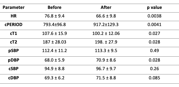
Introduction: Facial cooling is a simple way to stimulate trigeminal nerve receptors and evoke complex hemodynamic response that includes activation of both parasympathetic and sympathetic nervous system[1]. We ought to investigate a very early response to facial cooling in young heathy adults using pulse wave analysis (PWA), that enables to examine hemodynamic changes in aorta during cardiac cycle and its implications affecting systolic function of the heart. Objectives: To investigate an early hemodynamic response to facial cooling in young, healthy adults. Method: Study was conducted according to bioethical standards and Declaration of Helsinki. 15 volunteers with no cardiovascular diseases aged 18 to 25 (males: 10) were enrolled into the study. After 5 min of rest in supine position, brachial BP was measured and pulse waveform was recorded using applanation tonometry. Then participant was exposed to 15-second facial cooling, which was followed by second pulse waveform recording and brachial BP measurement. The following parameters were obtained: heart rate (HR), cPERIOD (central pulse period) peripheral and central systolic pressure (SP) and diastolic pressure (DP), C_T1 (the time from the beginning of pulse wave to systolic peak) and C_T2 (the time from the beginning of pulse wave to the reflected wave return peak). Results: FC caused decrease in HR and elongation of cPERIOD. There was a significant decrease in cT1, while cT2 increased. There was no significant change in peripheral and central SBP, as well as in the central DBP. However, there was a statistically significant increase in peripheral DBP. Statistical analysis was performed using R version 3.6.3 [2]. Due to insufficient sample size, it was impossible to accept the normal distribution as an approximation of our results’ distribution. Consequently, we conducted the analysis using Wilcoxon test. Conclusions: Despite a major rise in parasympathetic activity causing decrease in HR and elongation of cPERIOD, there was an increase in left ventricular systole velocity, manifested as shortened cT1. We hypothesized that it might be an effect of cPERIOD prolongation, consequent increase in end-diastolic volume (preload) and left ventricular systolic force and velocity. Another factor that may impact velocity of systole is decreased afterload, commonly defined as pressure in aorta. We didn’t obtain any significant change in cSBP nor cDBP, however there was a notable prolongation of cT2, reflecting increase of aortic compliance. It resulted in delay of reflected wave peak return and decrease of the pressure load to be overcome by the left ventricle, without decreasing maximum pressure in aorta. Our hypothesis is that during sympathetic activation ascending aorta acts differently than peripheral vessels.
Physiology 2021 (2021) Proc Physiol Soc 48, PC005
Poster Communications: Autonomic nervous system response to facial cooling increases velocity of left ventricle contraction and aortic compliance measured by pulse wave analysis.
Agata Janczak1, Beniamin Abramczyk2
1 Department of Internal Medicine, Hypertension and Vascular Diseases, Medical University of Warsaw, Warsaw, Poland 2 Institute of Evolutionary Biology, Faculty of Biology, Biological and Chemical Research Centre, University of Warsaw, Warsaw, Poland
View other abstracts by:
Where applicable, experiments conform with Society ethical requirements.