
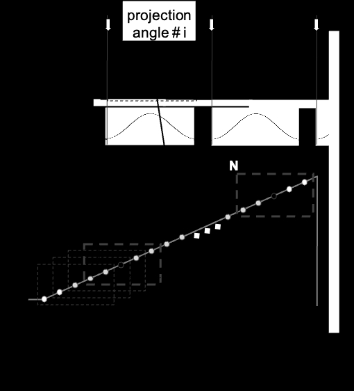
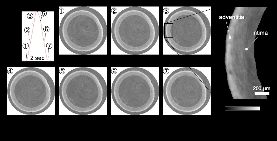
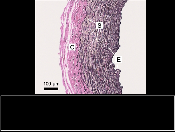
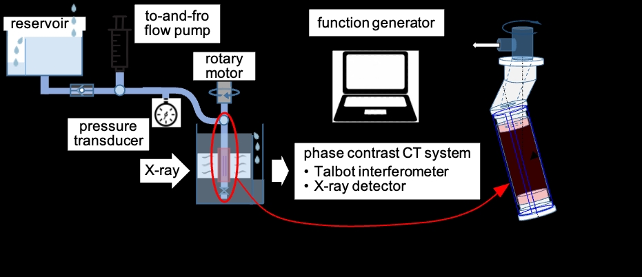
X-ray phase computed tomography (PCT) is highly sensitive in detecting density differences between structure components of biological soft tissue and well-suited to visualize the density-based tissue structure. Recently, PCT was successfully used to visualize repeated motions of soft tissue [1]. In this study, we applied PCT for visualizing arteries under pulsatile pressure condition and evaluated its feasibility in microstructure measurement of the entire circumferential arterial wall. A porcine carotid artery, purchased from a slaughterhouse, was measured by PCT at SPring-8 synchrotron radiation facility. The arterial segment was closed at one end by a plug. The other end was connected to a T-shape connector that was attached to a rotational stage on one side and connected on the other side to an overflow reservoir via a polyethylene tubing with an in-line check valve (Fig. 1). The tubing was filled with physiological saline. To induce pulsatile pressure waves, a programmable syringe pump was used to move the saline through a branch tubing in a to-and-fro motion over 2 s with 1-s rest insertion. The arterial segment was axially stretched by 30% and placed in a saline-filled container. A PCT system used a Talbot grating interferometer for phase measurement. The grating interferometer was composed of a phase grating (G1) and an absorption grating (G2), both with the pitch size of 2.4 μm, and optimized for the 20-keV X-ray. A fiber coupled sCMOS camera equipped with a beam monitor and a P43 phosphor screen was used as an X-ray detector. The effective pixel size was 4.47 μm. Schematic drawings of the image acquisition procedure are shown in Fig. 2. Phase retrieval was achieved by 5-step fringe scan, where the G2 mounted on a piezo stage was scanned for phase stepping with sequential saw-tooth wave signals from a function generator. The trigger signal from the syringe pump generated at the end of each pulsatile pressure was used to synchronize the saw-tooth wave generation as well as the projection angle increment. Images were acquired with 40-ms exposure at 50-ms intervals, yielding 60 images during each pressure pulsation or equivalently at each angular position of sample rotation over 180° in 0.2° steps. Cross-sectional tomographic images of the artery were shown at seven time points in a single pressure pulsation in Fig. 3, with the magnified image in the rectangle. The density resolution was estimated to be 2.5 mg/cm3. When the pressure was increased from 70 to 90 mmHg, the equivalent inner diameter calculated from the luminal area increased by 2.5% from 2.81 cm to 2.88 cm, and the wall volume of 3.8-mm long segment decreased by 0.6%. The outer layer is higher in density than the inner layer; the latter presented light- and dark-gray bands (i.e., different density bands). Elastica van Gieson staining confirmed that the image contrast in PCT was attributed to different components of vascular wall (Fig. 4). The present PCT has the potential to promote better understanding of arterial wall dynamics as well as risks of arterial rupture and dissection.
Physiology 2021 (2021) Proc Physiol Soc 48, PC089
Poster Communications: Phase contrast X-ray CT for imaging of the entire circumferential structure of arteries under pulsatile pressure condition
Takeshi Matsumoto1, Hiroyuki Tachibana2, Masato Hoshino3
1 Tokushima University, Tokushima, Japan 2 Kawasaki University of Medical Welfare, Kurashiki, Japan 3 SPring-8, Sayo, Japan
View other abstracts by:
Where applicable, experiments conform with Society ethical requirements.