
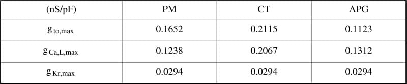
We constructed a biophysically detailed and anatomically accurate computer model of human atria that incorporates both structural and electrophysiological inhomogeneities. The 3D geometry was extracted from the Visible Female dataset. The sinoatrial node (SAN) and atria, including crista terminalis (CT), pectinate muscles (PM), appendages (APG) and Bachmann’s bundle (BB) were segmented using interactive deformable meshes. Fibre orientation in the bundles was set to local longitudinal direction. Ionic models based on Courtemanche et al. (1998) were used for describing cellular electrophysiology of all tissue types. The ionic channel conductances of Ito, ICa,L and IKr for PM, CT and BB were modified (Tab) to reproduce the action potential duration distributions of Feng et al. (1998). Pacemaker activity in the SAN was reproduced by removing IK1, including If and ICa,T, and modifying further channel properties using formulations of Zhang et al. (2000). Anisotropic propagation was computed with a monodomain approach using the finite element method. The excitation was first initiated in the centre of the SAN (Fig 1A), then conducted preferentially towards the atrioventricular region via the CT (Fig 1B) and afterwards spread over the right atrium along the PM (Fig 1C). Earliest activation of the left atrium was in the region of BB and excitation spread over to the appendage (Fig 1D). The conduction velocities were 60 cm/s for atrium, 120 cm/s for CT, 140 cm/s for PM, and 110 cm/s for BB at a frequency of 63 bpm. The simulation demonstrates the important role of anatomical and electrophysiological heterogeneity for a realistic propagation of excitation in the atrium. The preferential conduction towards CT and along PM is consistent with clinical endocardial mapping.
University of Oxford (2004) J Physiol 561P, PC26
Communications: 3D ANATOMICAL AND ELECTROPHYSIOLOGICAL MODEL OF HUMAN SINOATRIAL NODE AND ATRIA
Seemann,Gunnar ; Sachse,Frank B; Doessel,Olaf ; Holden,Arun V; Boyett,Mark R; Zhang,Henggui ;
1. Institute of Biomedical Engineering, University Karlsruhe (TH), Karlsruhe, Germany. 2. Nora Eccles Harrison Cardiovascular Research and Training Institute, University of Utah, Salt Lake City, UT, USA. 3. School of Biomedical Science, University of Leeds, Leeds, LS2 9JT, United Kingdom. 4. Dept. of Biological Physics, UMIST, Manchester, M60 1QD, United Kingdom.
View other abstracts by:
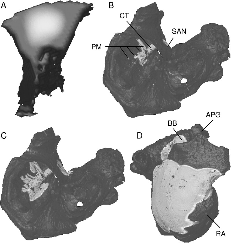
Figure 1: Gray-coded transmembrane voltage during excitation propagation in human atria. (A) SAN visualised only: Activation starts in the centre of the SAN; (B+C) View trough valves: Excitation spreads along CT and PM; (D) Lateral view: Right atrium and left via BB gets activated.
Where applicable, experiments conform with Society ethical requirements.