
Morgagni (1761): “Coronary arteries: three, […] sometimes four […]” [1] The human coronary tree is commonly portrayed as having two roots only: left and right coronary arteries (LCA, RCA; respectively). However, a third coronary artery (TCA) originating from the right coronary sinus has been observed regularly by anatomists in dedicated necroscopies, as early as the 18th century. Using magnetic resonance imaging (MRI) to reconstruct 3D cardiac tissue-architecture and the coronary vasculature, we identified TCAs in human and New Zealand White rabbit (NZ-white), and reconstructed their configuration. Hearts from 9 NZ-white rabbits (female) and 6 human cadavers were excised and fixed, gadolinium-treated, and agar-embedded for high-resolution MRI, followed by 3D reconstruction [2,3]. TCAs were present in all human and rabbit hearts imaged, originating either from independent aortic ostia (5/9 rabbits; 2/6 humans) or an ostium shared with RCA (but not as a branch of the RCA). TCA cross-sectional area was 26.7±10.1% of RCA in human, and 15.3±6.0% in rabbit (mean±SD). In all-but-one-case, TCA originated ventrally of RCA and progressed towards the Conus arteriosus of the right ventricular outflow tract (RVOT; 8/9 rabbits, 3/6 humans; progression not traceable in 1 human due to resolution, and in 2 humans because of restricted size of the overall tissue sample available). In one rabbit, a TCA with separate ostium originated dorsally of the RCA and progressed towards the Crista terminalis (forming the sinus node artery). Additionally, a fourth coronary vessel was found, which proceeded towards the aortic wall (7/9 rabbits; 3/6 humans; not traceable in 1 human due to resolution, and in 2 humans because of tissue sample quality). This was either an early branch of the TCA (5/7 rabbits, 2/3 humans), or arose from an ostium common with (1/7 rabbits, 1/3 humans) or independent of the TCA (1/7 rabbits). A TCA originating from the right coronary sinus is common, not only in human but also in rabbit, suggesting the latter may be suitable for related functional research. Conus arteriosus TCAs feed the RVOT and support blood supply to the early aortic wall. By and large, diagnostic and therapeutic interventions that target coronary vasculature do not consider the possibility of multiple ostia in the right coronary sinus. Better awareness of coronary tree structure and function may be beneficial for these procedures and, potentially, for understanding the mechanisms underlying RVOT rhythm disturbances and/or aortic root dysfunction.
University of Manchester (2010) Proc Physiol Soc 19, C56
Oral Communications: 3D histoanatomical reconstruction of the heart: rediscovering the Third Coronary Artery
R. A. Burton1, T. A. Quinn1, M. J. Bishop2, P. W. Hales3, C. Bollensdorff1, J. E. Schneider3, P. Kohl1
1. Physiology, Anatomy and Genetics, University of Oxford, Oxford, Oxfordshire, United Kingdom. 2. Computing Laboratory, University of Oxford, Oxford, United Kingdom. 3. BHF Experimental MR Unit, Department of Cardiovascular Medicine, University of Oxford, Oxford, United Kingdom.
View other abstracts by:
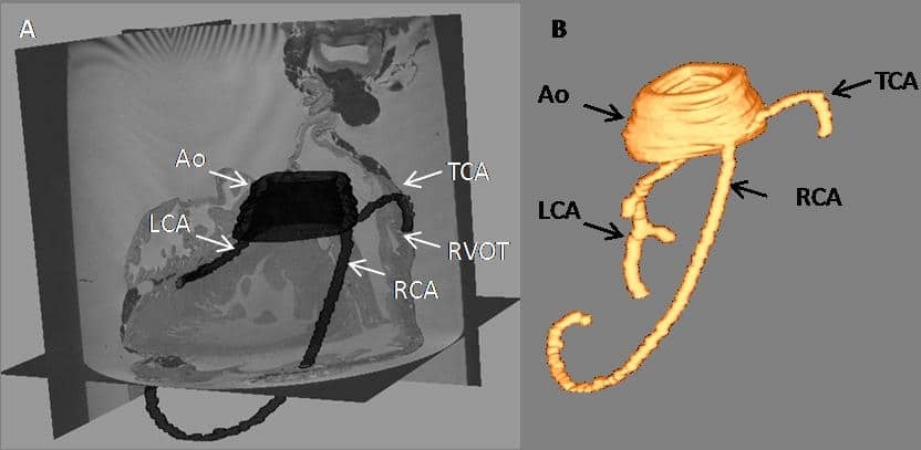
Figure 1: 3D dorso-lateral view of segmented coronary vessels in a rabbit heart. A: Segmented aortic root (Ao), left, right and third (Conus arteriosus) coronary arteries (LCA, RCA, TCA respectively). Here the segmented vessels are superimposed with 2D MRI image stacks of the cardiac tissue, using Seg3D software for visualisation. The TCA originates ventrally of the RCA, proceeding towards the right ventricular outflow tract (RVOT). B: Segmented vessels generated using Taratula and Meshalyzer for visualisation.
Where applicable, experiments conform with Society ethical requirements.