
Stretch of single cardiomyocytes acutely raises Ca2+ spark rate via a microtubule mediated mechanism of unknown nature [1,2]. Prior confocal and immuno-gold transmission electron microscopy suggested that microtubules co-localise with sarcoplasmic reticulum ryanodine receptors [3]. However, antibody-dependence and 2D nature of these techniques makes quantitative assessment of co-localisation difficult. Here, we use electron tomography (5 nm resolution) [4] to study the 3D spatial interrelation of microtubules, sarcoplasmic reticulum, and T-tubular system. Adult rat ventricular myocytes were chemically fixed, post-fixed in OsO4, dehydrated and embedded in resin. Sections (250 nm) were imaged with a 300kV electron microscope (Tecnai F30). Dual-axis tilt-series (1° steps, ±60° per axis) were acquired, aligned and combined to reconstruct 3D volumes (tomograms) from five different cells. Tomograms were then processed to generate 3D models used to quantify spatial relationship of microtubules, sarcoplasmic reticulum and T-tubular membranes (Figure 1). Microtubules (tubular structures with a diameter of 24 nm) regularly traverse the sarcoplasmic reticulum–T-tubular membrane complex which contains the cytoplasmic domain of ryanodine receptors. Microtubules approach sarcoplasmic reticulum and T-tubular membranes to within 7 nm and 13 nm, respectively, suggesting spatial proximity that is close enough to support mechanical interaction. Thus, electron tomography allows quantitative assessment of the 3D interrelation of microtubules, sarcoplasmic reticulum and T-tubules in adult rat ventricular cardiomyocytes. Microtubules are found in very close proximity to sarcoplasmic reticulum and T-tubular membranes, potentially supporting mechanical transmission of information from the extracellular environment to the sarcoplasmic reticulum–T-tubular complex. Thus, microtubules could contribute to the acute mechanical modulation of cellular Ca2+ handling.
University of Cambridge (2008) Proc Physiol Soc 11, PC18
Poster Communications: 3D reconstruction of microtubules, sarcoplasmic reticulum, and T-tubular membrane interrelation in rat ventricular myocytes using electron tomography
P. Camelliti1, F. Mason1, M. K. Morphew2, A. Hoenger2, P. Kohl1
1. Department of Physiology, Anatomy & Genetics, University of Oxford, Oxford, United Kingdom. 2. Department of Molecular, Cellular & Developmental Biology, University of Colorado, Boulder, Colorado, USA.
View other abstracts by:
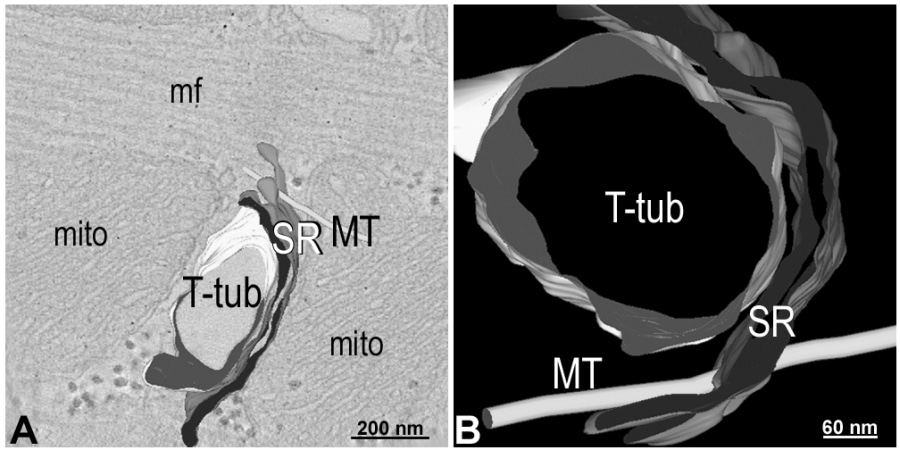
Figure: 1: A) Electron tomogram section and 3D model of a microtubule (MT) near sarcoplasmic reticulum (SR) and T-tubular (T-tub) membranes. B) Zoomed and rotated model showing details of spatial interrelation. mito: mitochondria; mf: myofilaments.
Where applicable, experiments conform with Society ethical requirements.