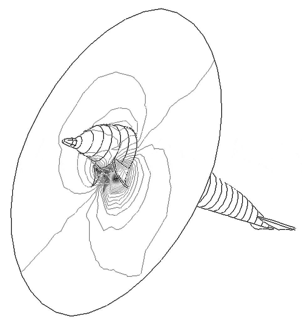
Diving in mammals is an exciting physiological challenge in that a number of cardiac and vascular reflexes are initiated to adjust the cardiac output to the reduced oxygen availability. However, quantification of electric cardiac activity during submergence in seawater is hampered by several technical problems, including signal attenuation, but also, for mammals other than man it is necessary to ascertain the optimal position for each electrode placement. To answer both questions we modelled a dolphin body, its heart, its skin and the surrounding seawater.
It is assumed that the heart polarization vector changes slowly so that electromagnetic effects can be disregarded and the electrodynamic problem can be solved as a succession of electrostatic problems. Under this assumption, each electropotential distribution can be obtained through the solution of the Laplace equation. The method chosen for the solution of Laplace’s equations was the finite element method (FEM), which allowed the electrostatic field boundary conditions between dolphin body, skin and seawater, without the need of mathematical enforcement. In order to solve the numerical problem, the dolphin body and fins have been discretized and covered with a skin (Behrmann, 1997). Then, the discrete model was submerged in a seawater mesh. Finally, the model was completed by incorporation of tissue (muscle and skin) resistivities (Hyttinen et al. 1997). These values were measured in the laboratory with a four-point probe on dead specimens.
Assuming that the heart acts as a dipole, the limit in the seawater around the dolphin was taken at a distance far enough to avoid the influence of boundary conditions in the electropotential distribution within the dolphin. The dipole intensity and direction within the dolphin heart is assumed from comparison with the human heart. Care was taken with the ventricular repolarization, the T wave, which appears as a deep downward deflection in a dolphin ECG.
The simulated ECG, without the seawater, was validated by comparing the modelled ECG with the records obtained in captive dolphins, the results showing a very good agreement both in shape and intensity. After this validation, seawater was added to the model and its influence on the signal levels and distribution was analysed (Fig. 1).

