
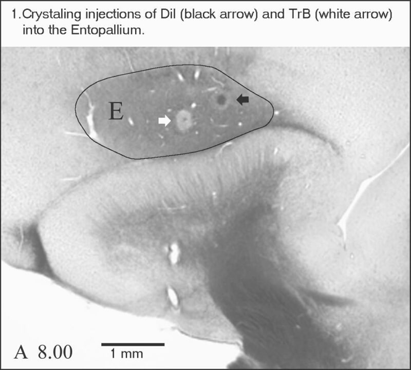
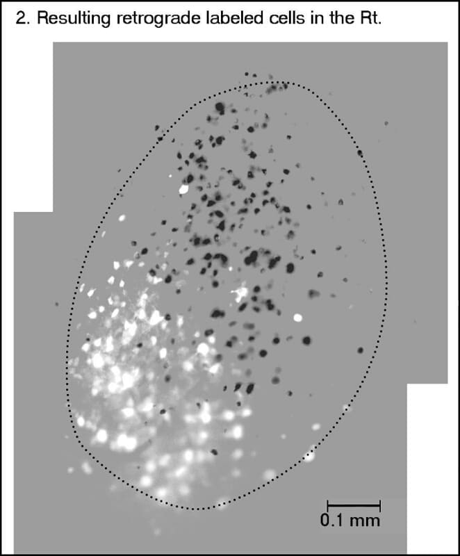
The tecto-fugal pathway is the major ascending visual pathway in non-mammalian vertebrates. In pigeons, most of the retinal axons target the optic tectum in a strictly topographical manner. The tecto-fugal projections arise exclusively from several different kinds of layer 13 tectal ganglion cells, which in turn target the different subdivisions of the thalamic nucleus rotundus (Rt) in an interdigitating non-retinotopic fashion (Marin et al. 2003). The cells in the Rt innervate the entopallium (E), a nuclear area that lies in the pallial telencephalon. Previous studies in zebra finch have shown that the rotundus-entopallial projections have a zonal topography, so that the rostro-caudal order of the rotundal subdivisions is preserved through the projection (Lavergheta & Shimizu, 2003; Krutszfeldt & Wild, 2004). However, the detailed organization of the projection from each rotundal subdivision to the E has yet to be described. We have investigated this organization performing pin-point single and double injections of small crystals of the retrograde tracers Dil and True Blue into a given zone of the E of adult pigeons, using a specially designed solid microinjector apparatus (Marin et al. 2001). Pigeons were anesthetized with a mixture of Ketamine (40 mg/kg) and Xilazine (12 mg/kg) injected intramusculary in the pectoral muscles. Anaesthesia was maintained with supplementary doses every 2 h. The E was sterotaxically reached through a small craneotomy exposing the dorsal telencephalon. Local incisions and pressure points were treated with anaesthesic ointment. After recuperative surgery and a survival period of 3-5 days animals were deeply anaesthetized and perfused via the aorta with 4% paraformadehyde. The brains were excised and processed according to standard procedures. Single injections into restricted loci of the E resulted in the labeling of a well-defined cluster of cells in the Rt. These clusters are mainly confined within the limits of a given rotundal subdivision. Double injections of DiI and True Blue located near one another in the same zone of E (figure 1) resulted in two close but clearly discernible clusters of retrograde labeled cells in the Rt (figure 2, dark cells labeled with Dil and white cells labeled with True Blue). These clusters appear to be confined to a single subdivision (in the figure 2, posterior subdivision), and the distance between them is proportional to the distance between the crystals in the E. Double-labeled cells were very scant, and located only in the overlapping periphery of the clusters. These results strongly suggest there is a broad, but consistent point-to-point topographical arrangement in the efferent projection of each rotundal subdivision to its corresponding entopallial zone. Thus, the activity of a given ectostriatal region could be modulated by the activity of a specific distributed ensemble of tectal ganglion cells via an intermediate cluster of rotundal neurons.
King's College London (2005) J Physiol 565P, C116
Communications: A Detailed Study of the Organization of the Rotundo-Entopallial Projections in the Pigeon (Columba livia)
Mpodozis, Jorge ; Fredes, Felipe ; Sentis, Elisa ; Tapia, Sebastian ; Marin, Gonzalo ; Letelier, Juan Carlos ; Maturana, Humberto ;
1. Departamento de Biologia, Universidad de Chile, Facultad de Ciencias, Santiago, Chile.
View other abstracts by:
Where applicable, experiments conform with Society ethical requirements.