
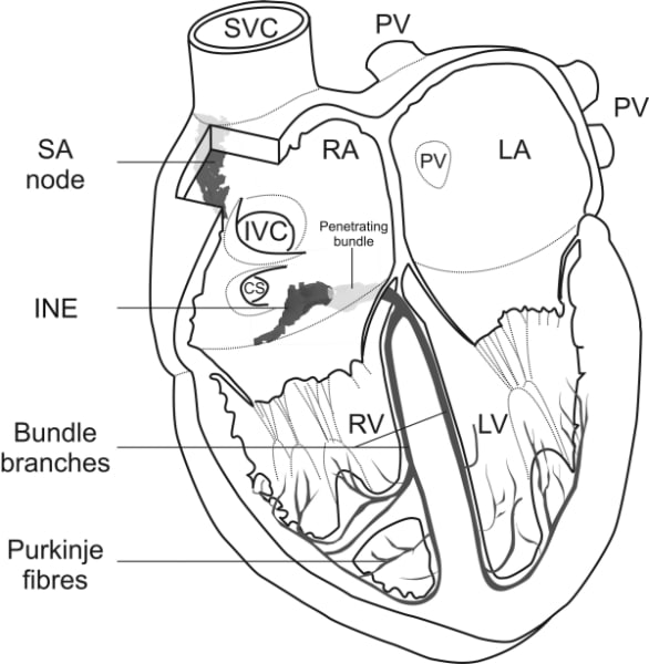
Based on electrophysiology, histology, quantitative PCR, in situ hybridisation and computer modelling, we have constructed a ‘model’ of the rabbit atrioventricular node: a tract of neurofilament- (NF-) positive nodal tissue – the ‘penetrating bundle’ – plunges through the fibrous layer insulating the atria from the ventricles and emerges in the ventricles as the His bundle (Fig. 1). There are two inputs into the penetrating bundle: slow and fast pathways. The slow pathway is most likely the ‘inferior nodal extension’ (INE), a ~5 mm tract of small dispersed NF-positive nodal tissue. At its start it is connected to atrial tissue and at its end it is continuous with the penetrating bundle, but in the middle it may be insulated by a vein. Just like the sinus node and different to the working myocardium, the INE lacks expression of Cx43 and Nav1.5 and expresses HCN4 and Cav1.3; the result is expected to be a slowly conducting ‘Ca2+-dependent’ action potential. We suggest that the INE is made up of N cells (cells, identified using electrophysiology, with these characteristics). Paradoxically, the penetrating bundle (including the ‘compact node’), although NF-positive, expresses some Cx43 and Nav1.5 and less HCN4 and we suggest that it is made up of NH cells (transitional Nodal-His cells as identified using electrophysiology). The fast pathway is most likely made up of transitional cells (NF-negative like atrial cells, but small and dispersed like nodal cells). Although it expresses Cx43 like the working myocardium, it may express Nav1.5 at a reduced level compared to the working myocardium. It is likely, therefore, that the fast pathway is made up of AN cells (transitional Atrial-Nodal cells as identified using electrophysiology). We have tested this ‘model’ of the atrioventricular node using computer modelling and we are able to simulate many of the physiological and pathophysiological behaviours of the atrioventricular node. For example, a model of a string of atrial cells connected via parallel strings of N cells (slow pathway) and AN cells (fast pathway) to a string of NH cells (penetrating bundle) – each cell type has its own biophysically detailed action potential – is able to simulate atrioventricular nodal reentrant tachycardia. Fig. 1. Diagram of heart incorporating detailed anatomical models of the sinoatrial and atrioventricular nodes.
University of Manchester (2007) Proc Physiol Soc 8, SA11
Research Symposium: A ‘model’ of the atrioventricular node
M. R. Boyett1, I. D. Greener1, J. Li1, S. Inada1, J. O. Tellez1, R. Billeter2, H. Zhang1, J. C. Hancox3, I. R. Efimov4, H. Dobrzynski1
1. Cardiovasculoar and Endocrine Science, Manchester University, Manchester, United Kingdom. 2. Nottingham university, Nottingham, United Kingdom. 3. Bristol university, Bristol, United Kingdom. 4. Washington University, St. Louis, MO, USA.
View other abstracts by:
Where applicable, experiments conform with Society ethical requirements.