
Haemodynamic forces occur naturally at branches of medium and large arteries and contribute to the development of atherosclerotic lesions. These forces modulate gene expression in the endothelium through complex mechanosensitive pathways. Low and oscillatory flow patterns promote a pro-atherogenic genotype, while high laminar flow activates athero-protective genes. To simulate pathophysiological, atherogenic blood flow patterns we have developed an in vitro perfusion system which allows exposure of endothelial cells grown in micro-slides to low or oscillatory flow patterns. Using this perfusion system we have examined the preconditioning effect of oscillatory flow on endothelial cells. Preconditioning human umbilical vein endothelial cells (HUVECs) with oscillatory flow enhanced their responses to inflammatory stimulation. Thus, subsequent exposure of HUVECs for 18-22 hours with the inflammatory cytokine TNFα (5 ng/ml) resulted in increased detection of pro-inflammatory and chemoattractant factors such as IL-8 and MCP-1, when compared to non-preconditioned cells (Table 1). When compared to static controls, we also demonstrated that preconditioning cells affected the distribution of the adhesion molecule ICAM-1, as determined by immunocytochemical staining. In an adhesion assay, preconditioning further affected the adherence of THP-1 monocytes to HUVECs. In summary, this system allows the assessment of responses of the vascular endothelium to inflammatory factors in a pathophysiologically-relevant setting. Priming of HUVECs with oscillatory flow altered their protein expression and functional profiles and rendered them more sensitive to subsequent treatment with TNFα. This in vitro model is amenable to further studies examining the effects of cigarette smoke toxicants on the vascular endothelium.
King's College London (2008) Proc Physiol Soc 13, C11
Oral Communications: A novel in vitro model for studying responses of endothelial cells under physiological flow conditions
N. Cockcroft1, R. Zantl2, T. Oke1, F. Cunningham1, E. Bishop1, I. Fearon1, M. D. Gaca1
1. Group R & D, British American Tobacco, Southampton, United Kingdom. 2. ibidi GmbH, Munich, Germany.
View other abstracts by:
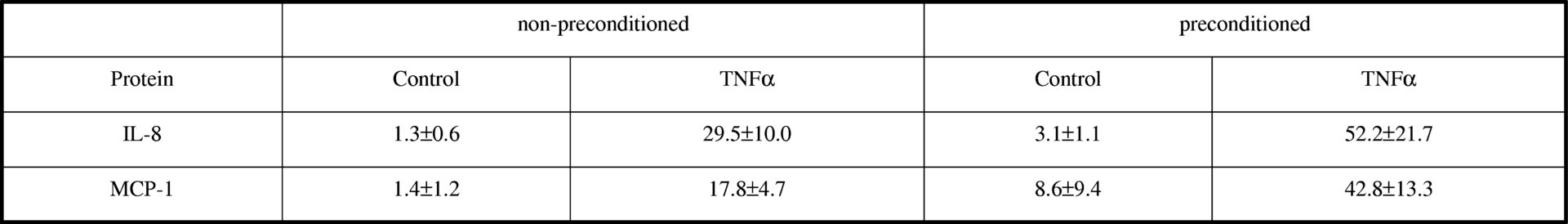
Table 1. Concentrations of inflammatory and chemoattractant proteins in media following exposure of HUVECs for 18-22 hours to 5 ng/ml TNFα. Data were obtained in cells cultured in static conditions (non-preconditioned) or preconditioned with oscillatory flow. Protein levels were determined by multiplex electrochemiluminescence detection using the MesoScale Discovery (MSD) platform. All concentrations are μg/ml. Data are means ± S.D. (n = 5-7 in each case).
Where applicable, experiments conform with Society ethical requirements.