
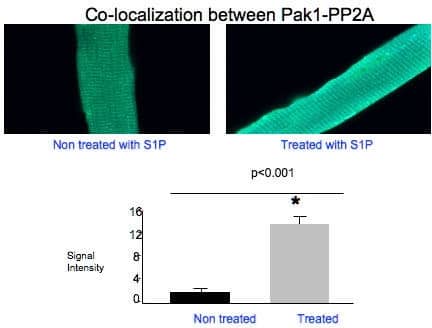
Sphingosine 1-phosphate (S1P) is bioactive lipid derived from metabolism of sphingomyelin that has been implicated in regulation of many cellular functions including ion channel activities (Hla, 2000; Pyne, 2000). To determine the effect of S1P on L-type calcium current (ICa,L) at basal and β-adrenergic stimulation conditions and underlying intracellular signalling pathways in rat ventricular cardiac myocytes, Western blot, immunocytochemistry and voltage patch clamping were used. The results are presented as mean ± SEM. Analysis was performed using the paired or un-paired Student’s t test as appropriate and significance was accepted at p 0.05). The effect of S1P on ICa,L was then studied in the presence of β-adrenergic stimulation by perfusion of 100 nM isoproterenol (ISO), a β-adrenergic receptor agonist. ICa,L was significantly enhanced in the presence of 100 nM ISO, the peak current density at 0 mV increased by 83.27 ± 2.8% from -3.44 ± 0.07 at the basal condition to -20.57 ± 0.76 pA/pF in the presence of ISO (p < 0.01, n = 7). ISO had a significant positive effect on ICa,L. Additional 100 nM S1P significantly blunt effect of ISO on ICa,L (in the presence of ISO), the peak current density at 0 mV increased by 68.99 ± 3.4% from -3.44 ± 0.07 to -11.1 ± 1.80 pA/pF (p < 0.01, n = 7). Our immunocytochemistry results also showed the co-localization between PAK1 (p21 activated kinase) – PP2A (protein phosphatase 2A) (n=25, p<0.001). Western blotting showed that the S1P1 receptor expressed in the cells whereas no visualization of S1P2 and S1P3 signals were observed. In conclusion, in rat ventricular myocytes, S1P does not affect the basal activity of L-type Ca channels but reversed the effect of the β-adrenergic agonist ISO on this channel. This effect is probably through PAK1-PP2A-mediated signalling pathway via S1P1 receptor.
University of Manchester (2007) Proc Physiol Soc 8, PC6
Poster Communications: A novel mechanism of sphingosine 1-phosphate signalling in regulation of L-type Ca2+ channel activity in cardiac myocytes
E. Eroume A Egom1, Y. Li1, H. Musa1, M. Lei1
1. Division of Cardiovascular and Endocrine Sciences, University of Manchester, Manchester, United Kingdom.
View other abstracts by:
Where applicable, experiments conform with Society ethical requirements.