
Langendorff mode of heart perfusion is widely used in biomedical research. It is a constant retrograde perfusion with unnatural pressure in four chambers, which causes abnormal heart conditions. For example, rabbit heart rate in vivo is 205~220/min at rest, whereas the heart rate of Langendorff perfused rabbit heart is 120~150/min (Döring & Dehnert, 1988). Furthermore, the preload of left ventricle in Langendorff perfused heart is highly increased as the aortic valve is unable to work as normal, which may cause acute congestive heart failure. To investigate arrhythmia in the isolated hearts from rabbit and rat, we have developed a physiological mode of heart perfusion in vitro that simulated the working heart in vivo as shown in Figure 1. In this system, the regular flow through the heart was maintained, and each chamber was automatically controlled and filled with changing volume and pressure as usual. As a result, normal heart rate in vivo could be kept in vitro for more than 24 hours. The pressure in each chamber was adjustable and controllable, which was useful to study those arrhythmia associated with stretch and/or pressure. To investigate the role of autonomic nerve system in atrial fibrillation, the system was also used to carry out the experiments in vivo with intact autonomic innervation of the heart perfused physiologically with Tyrode solution as shown in Figure 2. Based on characteristics of the system, this mode is valuable to functional studies on arrhythmia that is a systemic disorder at organ level and highly related to the pressure in heart chamber.
University College Dublin (2009) Proc Physiol Soc 15, PC28
Poster Communications: A physiological mode of heart perfusion in vivo and in vitro
G. Hao1, Y. Song1, M. Boyett1, X. Yang2, Y. Du2, A. Luo3, M. Tang3, Z. Shui1,2
1. Cardiovascular Research Group, University of Manchester, Manchester M13 9NT, United Kingdom. 2. Departments of Cardiovascular Surgery and Cardiology, Union Hospital, Wuhan, China. 3. Department of Physiology, Tongji Medical College, Huazhong University of Science and Technology, Wuhan, China.
View other abstracts by:
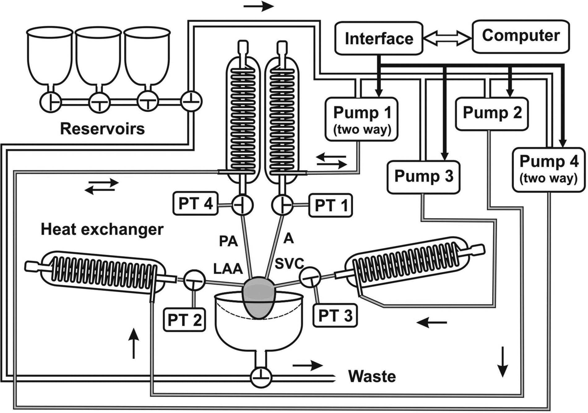
Figure 1. A physiological mode of heart perfusion in vitroPressure transducer (PT), pulmonary artery (PA), aorta (A), superior vena cava (SVC), left atrial appendage (LAA). Pulmonary veins and inferior vena cava were tied.

Figure 2. Physiological heart perfusion in vivo with intact autonomic innervationIn accordance with the Animals (Scientific Procedures) Act 1986 and the Guide for the Care and Use of Laboratory Animals (NIH Publication No. 85~23, 1985), anaesthesia was induced and maintained with Hypnoval (1 mg/kg, I.V.) and pentobarbitone (2 mg/5 min, I.V.) before and during dissection of the vagus nerves, and an overdose of pentobarbitone (60 mg, I.V.) was given before opening the thoracic cavity (André Ng et al. 2001). The descending aorta and inferior vena cava were connected, whereas SVC was tied.
Where applicable, experiments conform with Society ethical requirements.