
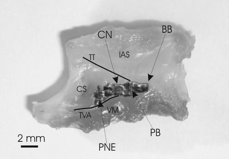
The atrioventricular (AV) node conduction axis is complex and heterogenous in terms of function and morphology. Functionally, there are dual (fast and slow) conduction pathways leading from the atrial muscle to the compact AV node (Wu, 1982). As well as normal conduction, the dual pathways are also implicated in AV node pacemaking and AV nodal reentrant tachycardia. Despite their functional importance, on an anatomical level, these pathways are poorly understood. To obtain a better understanding of the structure of the AV node, we are constructing a 3-D model of the region of the AV node (the triangle of Koch). Adult New Zealand White rabbits were killed humanely and the AV node region isolated (n = 6; Fig. 1). 10 µm serial sections were cut perpendicular to the tricuspid valve (and the endocardium). 60 sections at 100 µm intervals were stained with Masson’s trichrome to show the histology. Adjacent sections were immunolabelled for neurofilament (a marker of the conduction system in the rabbit heart) and Cx43 (gap junction protein known to be present throughout much of the heart, but not in the AV node). Based on the histology and the immunolabelling, the distributions of different cell types (neurofilament-negative ventricular muscle, neurofilament-negative atrial muscle, neurofilament-positive conduction tissue, connective tissue) were identified in each of the 60 sections. Matlab software is being used to develop a 3-D model from these data. As an example, Fig. 1 shows the distribution of neurofilament-positive conduction tissue (dark grey) in the triangle of Koch (from left to right): posterior nodal extension (PNE), compact node (CN), penetrating bundle (PB), bundle branches (BB), interatrial septum (IAS), tendon of Todaro (TT), coronary sinus (CS), tricuspid valve annulus (TVA) and ventricular myocardium (VM). The posterior nodal extension corresponds to the position of the slow pathway. Cx43 immunolabelling was abundant in the working myocardium, but sparse or absent in the conduction tissue. Our data show two different tissue types in the AV node region: neurofilament-negative/Cx43-positive tissue (atrial and ventricular muscle) and neurofilament-positive/Cx43-negative tissue (conduction tissue).
University of Glasgow (2004) J Physiol 557P, C14
Communications: A three-dimensional model of the rabbit atrioventricular node conduction axis
I.D. Greener,H.Dobrzynski1,J.Li1,V.Nikolski,M. Yamamoto1, R. Billeter,I. Efimov and M.R. Boyett
I.D. Greener,H.Dobrzynski1,J.Li1,V.Nikolski,M. Yamamoto1, R. Billeter,I. Efimov and M.R. Boyett
View other abstracts by:
Where applicable, experiments conform with Society ethical requirements.