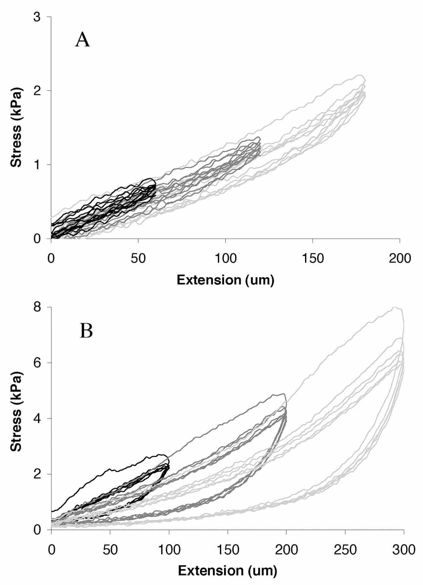
The mechanical behaviour of the diastolic heart reflects the passive properties of cardiac tissue, which have been reported to demonstrate ‘strain-softening’ (Emery et al. 1997). This phenomenon is characterised by a stiff stress-strain relationship on the first loading cycle; subsequent cycles at the same extension show a ‘softening’ of stress. If the tissue is then stretched to some greater extent, the first stress-strain curve of the new loading cycle follows the previous ‘softened’ locus. Subsequent cycles at the new extension again show ‘softening’ (Fig. 1B). In contrast to ‘stress relaxation’, strain softening is irreversible. It has been demonstrated in the pressure-volume behaviour of the 2,3-butanedione monoxime (BDM)-arrested rat heart (Emery et al. 1997) and by the shear behaviour of BDM-treated tissue blocks cut from the left-ventricular wall of the pig heart (Dokos et al. 2002). In both of these studies, the tissues were unperfused.
To determine whether strain softening occurs in well-oxygenated tissue, we subjected 10 superfused trabeculae, from the right ventricles of humanely killed rats, to repeated strain cycles of 5 %, 10 % and 15 % extension of muscle length. To ascertain the contribution of BDM, we tested half of the preparations first in the absence (-BDM) and then in the presence (+BDM) of 50 mM BDM. The other half experienced the converse order. Stress-strain data were subjected to repeated-measures, split-plot ANOVA (a = 0.05). As shown by the typical result in Fig. 1A (-BDM), we found no evidence of strain-softening – in either the presence or absence of BDM (data not shown). In fact, strain softening was observed (Fig. 1B; note difference of scales) only if trabeculae became non-viable (i.e. failed to respond to electrical stimulation). If strain-softening does not reflect deterioration of the health of the tissue, then it must reflect the different mode of deformation that we applied (linear elongation versus shear or volume expansion), or the different microstructure of trabeculae (axially aligned myocytes and perimysial collagen fibres (Hanley et al. 1999) vis-ˆ-vis the complex 3-D geometry of whole-hearts (Le Grice et al. 1995) or ventricular tissue blocks). We rule out a contribution from BDM.
This work was supported by the Marsden Fund and HRC (NZ).