
Ventricular failure is associated with remodelling of ion channels and intracellular Ca2+ dynamics. A model (Preibe & Beuckelmann, 1998) for normal and failing (remodelled IK1, INaCa, INaK, Ca2+-ATPase of the SR) heart cells was incorporated into a partial differential equation (PDE) tissue model to determine how the remodelling associated with heart failure can alter propagation patterns. We use the simple Euler method to integrate the PDE with a time step of 0.02 ms and a space step of 0.2 mm with a diffusion constant, D = 0.154 mm2/ms. Dynamic action potential duration (APD) restitution was obtained by pre-pacing the cell models 40 times to obtain the final diastolic interval (DI) and APD. Conduction velocity (CV) restitution was obtained by pacing the 1D spatial models for 5 s at a frequency of 1 Hz. CV of a 6th premature propagating wave was measured. Vulnerable window (VW) (Biktashev et al. 1998) in 1D was measured. The dynamic restitution curve for the failing case has a region of negative slope at high stimulation rate (small period) while is positive in the control case. The maximal magnitude of the slopes of both curves is < 1 and both the normal and failing models show alternans. At fast pacing rates electrical alternans were observed for a basic cycle length (BCL) of < 392 ms in control cell, and for BCL of < 665 ms in the failing cell. The CV for a solitary wave in the failing case is marginally greater than in the non-failing case (7%). The CV restitution is shifted upward in failing tissue. The measured vulnerable window (VW) for the tissue to re-entry is 5 ms for the control and 7 ms for the failing case. Heart failure increased vulnerability of the tissue to re-entry by 40% and shifted the timing of the window significantly. In conclusion, heart failure induced remodelling ion channel kinetics and intracellular Ca2+ handling has a significant impact on the rate dependent electrical action potential and conduction properties of excitation waves. It also increases vulnerability of the tissue to arrhythmogenesis.
University of Bristol (2005) J Physiol 567P, PC12
Poster Communications: Action potential duration, conduction velocity restitution, and vulnerability to re-entry in a computational model of human failing heart tissue
Kharche, Sanjay; Zhang, Henggui; Holden, Arun V;
1. Biomedical Sciences, University of Leeds, Leeds, United Kingdom. 2. Biological Physics Group, Department of Physics, UMIST, Manchester, United Kingdom.
View other abstracts by:
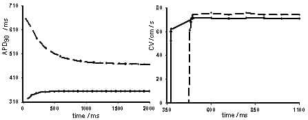
Figure 1. Restitution properties for normal (continuous dotted lines) and for failure (dash dotted lines) cell and tissue models. Left panel shows dynamic APD restitution for cell and right panel shows CV restitution curve for 1D tissue.
Where applicable, experiments conform with Society ethical requirements.