
The pulmonary veins (PV) are important in the initiation and maintenance of atrial fibrillation, which may be related to slow excitation-conduction in the PV (Nattel, 2002; Aldhoon et al. 2009). In this study, the essential factors for action potential conduction in the PV have been investigated with single myocytes isolated from rat left atrium (LA) and four PVs (left superior LSPV, left inferior LIPV, right superior RSPV and right inferior RIPV). Whole cell current clamp and voltage clamp were used to record action potential (AP) and the fast Na+ current (INa). Single cell real-time PCR was employed to measure the abundance of mRNAs for Nav1.5 and gap junction channel (Cx43), and the expression of Cx43 protein on cell membrane was located by immunofluorescence. The results showed that: [i] the maximum velocity of AP phase 0 upstroke was significantly faster (62±10.5 V/s) in LA myocytes than in all PV myocytes (Fig. 1 A and D); [ii] INa density was remarkably higher (16.4±2.9 pA/pF) in all PV myocytes (apart from RSPV myocytes) than in LA myocytes (Fig. 1 B, C and E); [iii] the relative abundance of Nav1.5 mRNA in the LSPV and LIPV myocytes was 1.7 times higher than in LA myocytes (Fig. 1F), whereas no difference in the Cx43 mRNA abundance between LA and PV myocytes was observed; and [iv] the majority of Cx43 expression in LA myocytes was located at the cell-ends in the intercalated disc, whereas the Cx43 expression in PV myocytes was distributed at both cell-ends and in the lateral cell membrane. It is surprising and interesting that INa density and the abundance of Nav1.5 mRNA in PV myocytes are inconsistent with the depolarization velocity of AP as compared with those in LA myocytes. In general, the conduction velocity is mainly associated with Na+ and gap junction channels as well as their functional balance, and INa is the large current that has sufficient reserve or redundancy for the fast depolarization. Therefore, it is concluded that the difference in the cellular distribution of Cx43 protein in PV myocytes is predominant to slow excitation-conduction as a result of anisotropic properties of the tissue more susceptible to atrial fibrillation.
University College Dublin (2009) Proc Physiol Soc 15, C123
Oral Communications: Action potential, sodium and gap junction channels in rat pulmonary vein myocytes
Y. Song1, G. Hao1, M. Boyett1, X. Yang2, Y. Du2, Z. Shui1,2
1. Cardiovascular Research Group, University of Manchester, Manchester M13 9NT, United Kingdom. 2. Departments of Cardiovascular Surgery and Cardiology, Union Hospital, Wuhan, China.
View other abstracts by:
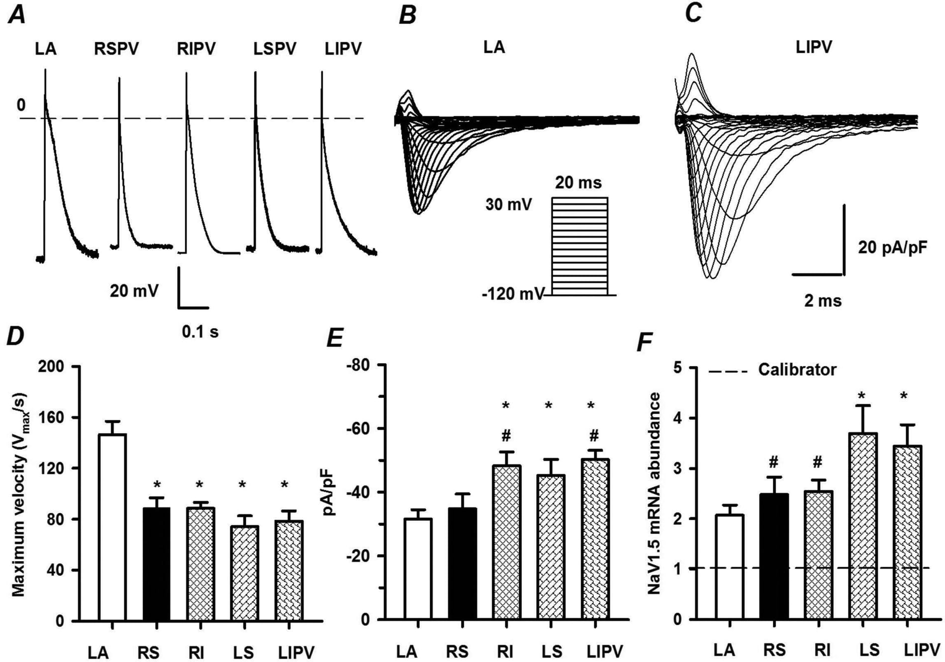
Figure 1. Action potential, INa and Nav1.5 mRNA in LA and PV myocytesA, recordings of action potentials from LA and PV myocytes. B and C, INa traces from a LA and a LIPV myocytes during voltage clamp. D, maximum velocity of phase 0 upstroke of AP in the various myocytes (n=10~19). E, peak density of INa (n=11~15). F, relative abundance of Nav1.5 mRNA (n=7). Significantly different * from LA myocytes and # from LSPV myocytes (P < 0.05, one-way ANOVA).
Where applicable, experiments conform with Society ethical requirements.